

Calpain-dependent clearance of the autophagy protein p62/SQSTM1 is a contributor to ΔPK oncolytic activity in melanoma
- PMID: 24553345
- PMCID: PMC3975656
- DOI: 10.1038/gt.2014.6
Calpain-dependent clearance of the autophagy protein p62/SQSTM1 is a contributor to ΔPK oncolytic activity in melanoma
Abstract
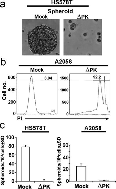
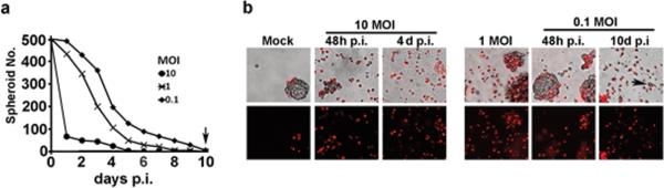
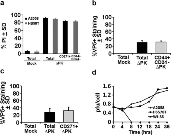
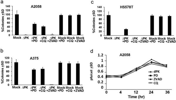
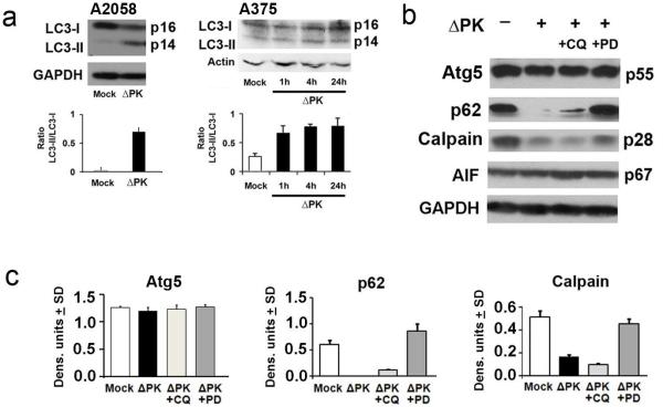
Oncolytic virotherapy is a promising strategy for reducing tumor burden through selective virus replication in rapidly proliferating cells. However, the lysis of slowly replicating cancer stem cells (CSCs), which maintain neoplastic clonality, is relatively modest and the potential contribution of programmed cell death pathways to oncolytic activity is still poorly understood. We show that the oncolytic virus ΔPK lyses CSC-enriched breast cancer and melanoma 3D spheroid cultures at low titers (0.1 pfu/cell) without resistance development and it inhibits the 3D growth potential (spheroids and agarose colonies) of melanoma and breast cancer cells. ΔPK induces calpain activation in both melanoma and breast cancer 3D cultures as determined by the loss of the p28 regulatory subunit, and 3D growth is restored by treatment with the calpain inhibitor PD150606. In melanoma, ΔPK infection also induces light chain 3 (LC3)-II accumulation and p62/SQSTM1 clearance, both markers of autophagy, and 3D growth is restored by treatment with the autophagy inhibitor chloroquine (CQ). However, expression of the autophagy-required protein Atg5 is not altered and CQ does not restore p62/SQSTM1 expression, suggesting that the CQ effect may be autophagy-independent. PD150606 restores expression of p62/SQSTM1 in ΔPK-infected melanoma cultures, suggesting that calpain activation induces anti-tumor activity through p62/SQSTM1 clearance.
Figures
Similar articles
-
ΔPK oncolytic activity includes modulation of the tumour cell milieu.J Gen Virol. 2016 Feb;97(2):496-508. doi: 10.1099/jgv.0.000353. Epub 2015 Nov 24. J Gen Virol. 2016. PMID: 26602205 Free PMC article.
-
The oncolytic virus ΔPK has multimodal anti-tumor activity.Pathog Dis. 2016 Jul;74(5):ftw050. doi: 10.1093/femspd/ftw050. Epub 2016 May 29. Pathog Dis. 2016. PMID: 27242376 Free PMC article. Review.
-
The HSV-2 mutant DeltaPK induces melanoma oncolysis through nonredundant death programs and associated with autophagy and pyroptosis proteins.Gene Ther. 2010 Mar;17(3):315-27. doi: 10.1038/gt.2009.126. Epub 2009 Oct 1. Gene Ther. 2010. PMID: 19798049 Free PMC article.
-
NF-κB Signaling Activation Induced by Chloroquine Requires Autophagosome, p62 Protein, and c-Jun N-terminal Kinase (JNK) Signaling and Promotes Tumor Cell Resistance.J Biol Chem. 2017 Feb 24;292(8):3379-3388. doi: 10.1074/jbc.M116.756536. Epub 2017 Jan 12. J Biol Chem. 2017. PMID: 28082672 Free PMC article.
-
Molecular Pathways: Mechanism of Action for Talimogene Laherparepvec, a New Oncolytic Virus Immunotherapy.Clin Cancer Res. 2016 Mar 1;22(5):1048-54. doi: 10.1158/1078-0432.CCR-15-2667. Epub 2015 Dec 30. Clin Cancer Res. 2016. PMID: 26719429 Review.
Cited by
-
Experimental diabetes exacerbates autophagic flux impairment during myocardial I/R injury through calpain-mediated cleavage of Atg5/LAMP2.J Cell Mol Med. 2023 Jan;27(2):232-245. doi: 10.1111/jcmm.17642. Epub 2022 Dec 23. J Cell Mol Med. 2023. PMID: 36562207 Free PMC article.
-
Three-dimensional tumor cell cultures employed in virotherapy research.Oncolytic Virother. 2018 Sep 5;7:79-93. doi: 10.2147/OV.S165479. eCollection 2018. Oncolytic Virother. 2018. PMID: 30234074 Free PMC article. Review.
-
Oncolytic virus as a cancer stem cell killer: progress and challenges.Stem Cell Investig. 2014 Dec 28;1:22. doi: 10.3978/j.issn.2306-9759.2014.12.02. eCollection 2014. Stem Cell Investig. 2014. PMID: 27358868 Free PMC article. Review.
-
ΔPK oncolytic activity includes modulation of the tumour cell milieu.J Gen Virol. 2016 Feb;97(2):496-508. doi: 10.1099/jgv.0.000353. Epub 2015 Nov 24. J Gen Virol. 2016. PMID: 26602205 Free PMC article.
-
The oncolytic virus ΔPK has multimodal anti-tumor activity.Pathog Dis. 2016 Jul;74(5):ftw050. doi: 10.1093/femspd/ftw050. Epub 2016 May 29. Pathog Dis. 2016. PMID: 27242376 Free PMC article. Review.
References
Publication types
MeSH terms
Substances
Grants and funding
LinkOut - more resources
Full Text Sources
Other Literature Sources
Medical
