. 2014 Feb 19;34(8):2989-3004.
doi: 10.1523/JNEUROSCI.2826-13.2014.

Extracellular calcium controls the expression of two different forms of ripple-like hippocampal oscillations

Affiliations

Extracellular calcium controls the expression of two different forms of ripple-like hippocampal oscillations

Paloma Aivar et al. J Neurosci. .

Abstract

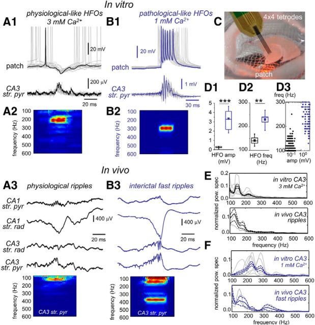
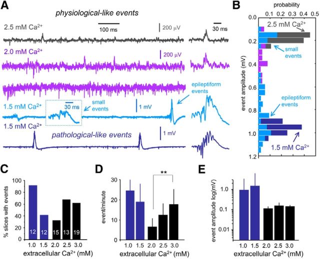
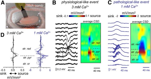
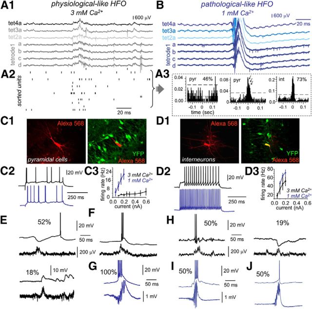
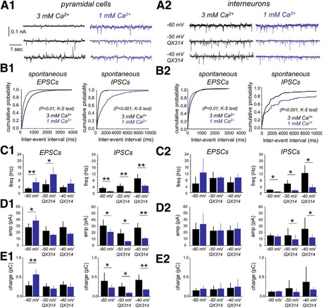
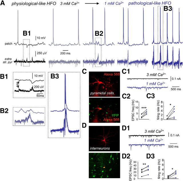
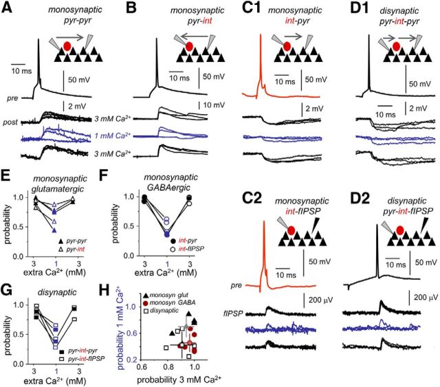
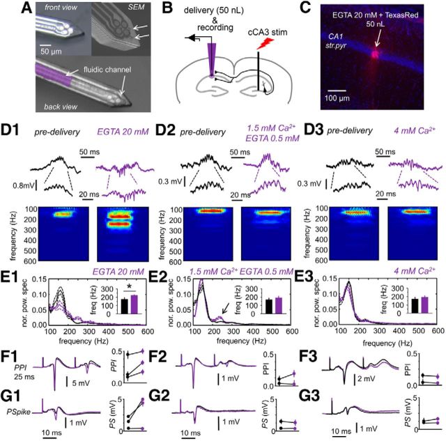
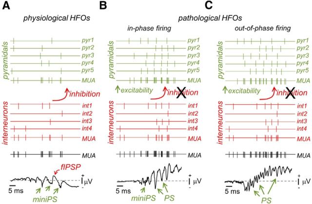
Hippocampal high-frequency oscillations (HFOs) are prominent in physiological and pathological conditions. During physiological ripples (100-200 Hz), few pyramidal cells fire together coordinated by rhythmic inhibitory potentials. In the epileptic hippocampus, fast ripples (>200 Hz) reflect population spikes (PSs) from clusters of bursting cells, but HFOs in the ripple and the fast ripple range are vastly intermixed. What is the meaning of this frequency range? What determines the expression of different HFOs? Here, we used different concentrations of Ca(2+) in a physiological range (1-3 mM) to record local field potentials and single cells in hippocampal slices from normal rats. Surprisingly, we found that this sole manipulation results in the emergence of two forms of HFOs reminiscent of ripples and fast ripples recorded in vivo from normal and epileptic rats, respectively. We scrutinized the cellular correlates and mechanisms underlying the emergence of these two forms of HFOs by combining multisite, single-cell and paired-cell recordings in slices prepared from a rat reporter line that facilitates identification of GABAergic cells. We found a major effect of extracellular Ca(2+) in modulating intrinsic excitability and disynaptic inhibition, two critical factors shaping network dynamics. Moreover, locally modulating the extracellular Ca(2+) concentration in an in vivo environment had a similar effect on disynaptic inhibition, pyramidal cell excitability, and ripple dynamics. Therefore, the HFO frequency band reflects a range of firing dynamics of hippocampal networks.

Keywords: drug delivery; fast ripples; high-frequency oscillations.


Figures

Figure 1.
Figure 1.
Physiological- and pathological-like HFOs. A1, Representative example of simultaneous extracellular and pyramidal cell patch recording during physiological-like HFOs in 3 mm Ca2+. A2, Time–frequency spectrum of the LFP recorded at the stratum pyramidale in A1. A3, In vivo recording of physiological ripples using 16ch silicon probes. One representative example is shown. For clarity sake, only data from relevant hippocampal strata at CA3 and CA1 are shown. str. pyr, Stratum pyramidale; str. rad, stratum radiatum. The time–frequency spectrum of the LFP recorded at the CA3 stratum pyramidale is shown at the bottom. B1, Example of simultaneous field potential and patch recording during pathological-like HFOs in 1 mm Ca2+. B2, Average time–frequency spectrum of the LFP recorded at the stratum pyramidale in B1. B3, Representative example of interictal fast ripples and time–frequency spectrum recorded from an epileptic rat in vivo. C, A modified chamber coupled to an upright microscope was used to record slices in interface conditions especially suitable for releasing spontaneous SPW–HFO events. A silicon-based tetrode (4×) was typically used to record LFPs and unit activity simultaneously with patch recording. Box plots of HFO amplitude (amp; D1) and HFO frequency (freq; D2) in 3 mm (black, n = 6 slices) and 1 mm (blue, n = 6 slices) Ca2+. D3, Group differences of HFO frequency and amplitude of each individual event clearly show clustering of data from 3 mm (black) and 1 mm (blue) Ca2+. E, Top, Normalized mean power spectrum for individual experiments (gray traces) and the grand average (black traces) of SPW–HFO events recorded in vitro at 3 mm Ca2+ (data from 6 slices from 6 rats). Bottom, Same for CA3 SPW–HFO events recorded in vivo from normal rats (data from 3 rats). Discontinuous lines reflect 95% confidence. F, Top, Normalized mean power spectrum of the individual experiments and the grand average of SPW–HFO events recorded in vitro at 1 mm Ca2+ (6 slices from 6 rats). Bottom, Same for CA3 SPW–HFO events recorded in vivo from epileptic rats (n = 3). **p < 0.01; ***p < 0.001.
Figure 2.
Figure 2.
Effect of extracellular Ca2+ on the features of spontaneous SPW–HFO events. A, Representative examples of the types of SWP-HFO events typically recorded in a range of extracellular Ca2+ concentration. Note the large multiunit activity recorded at 2 mm Ca2+ (purple) in slices not exhibiting spontaneous SPW–HFO events. Using 1.5 mm Ca2+ resulted in some slices exhibiting spontaneous SPW–HFO events of different amplitudes that were difficult to classify (light blue), whereas clear pathological SPW–HFO events were seen in others (dark blue). B, Amplitude histograms of all events detected in the representative slices shown in A. Note two clear peaks for small and large events detected in the slice at 1.5 mm Ca2+ (light blue, bottom trace). C, Dependence of the percentage of slices exhibiting spontaneous events on different extracellular Ca2+ concentration. Numbers indicate the total number of slices tested. D, Dependence of the rate of spontaneous SPW–HFO events on different extracellular Ca2+ concentration. **p < 0.0001. E, Dependence of event amplitude on different extracellular Ca2+ concentrations.
Figure 3.
Figure 3.
CSD analysis of spontaneous SPW events recorded in vitro at different Ca2+ concentrations. A, 16ch comb-like silicon probes were designed for these in vitro applications. B, Linear-oriented LFPs (100 μm interspacing) were obtained from CA3 in the somato-dendritic axis to evaluate CSD signals of SPW activity in 3 mm Ca2+. Traces show an individual event, whereas the CSD map depicts the average of many events. Average SPW-triggered CSD maps are color coded. C, Linear recordings and averaged SPW-triggered CSD map from a representative example in 1 mm Ca2+. D, Spatial profiles from SPW-triggered CSD signals from n = 5 slices (4 rats) in 3 mm Ca2+ (left, black) and in 1 mm Ca2+ (left, blue). str. pyr, Stratum pyramidale; str. rad, stratum radiatum; str. or., stratum oriens.
Figure 4.
Figure 4.
CA3 neuronal dynamics during physiological- and pathological-like HFOs. A1, Tetrode recordings were obtained to separate unit activity during physiological-like HFO in 3 mm Ca2+. A2, Firing increases of sorted units were observed to occur during physiological-like events. Units from data shown in A1. A3, Representative single-cell data. Many putative pyramidal units (46%) were inhibited during physiological-like HFO events, whereas most interneurons (73%) showed excitation. A minority of putative pyramidal cells fired burst during these events. B, Representative tetrode recordings obtained during pathological-like HFOs in 1 mm Ca2+. C1, A rat reporter line (VGAT–Venus A) known to express YFP in most hippocampal interneurons was used to patch from different cell types. Patched pyramidal cells were stained with Alexa Fluor 568 for subsequent colocalization analysis. C2, Representative suprathreshold responses of two different pyramidal cells recorded in 3 mm (black) and 1 mm (blue) Ca2+. C3, f/I curves from pyramidal cells recorded in 3 mm (black, n = 23) and 1 mm (blue, n = 9) Ca2+. D1, Alexa Fluor 568 and YFP colocalization allowed identification of GABAergic interneuron. D2, Responses of two different representative interneurons recorded in 3 mm (black) and 1 mm (blue) Ca2+ for current pulses of 0.2 nA. D3, f/I curves from interneurons recorded in 3 mm (black, n = 21) and 1 mm (blue, n = 11) Ca2+. E, Example traces of typical SPW-associated events recorded in pyramidal cells during physiological-like HFOs in 3 mm Ca2+. Many pyramidal cells exhibited clear SPW-associated inhibitory potentials (52%), whereas some others received excitatory inputs (18%). The remaining 17% cells did not show any SPW-associated response pattern. F, A minority of cells had bursting activity. G, Representative example of a pyramidal cell recorded during pathological-like HFO activity in 1 mm Ca2+. All recorded pyramidal cells were found to burst during HFO events. H, Representative examples of the SPW-associated responses of interneurons during physiological-like HFO in 3 mm Ca2+. I, SPW-associated high-frequency burst was recorded in 50% interneurons in 1 mm Ca2+. J, The remaining 50% of interneurons showed a variable participation typically contributing with single spikes in 1 mm Ca2+.
Figure 5.
Figure 5.
Spontaneous EPSC and IPSC dynamics in 3 mm and 1 mm Ca2+. A1, Spontaneous synaptic potentials were recorded from identified CA3 pyramidal cells at different membrane holding levels to detect excitatory (EPSCs) and inhibitory (IPSCs) components in 3 mm (n = 6 cells) and 1 mm (n = 6 cells) Ca2+. B1, Cumulative distribution of the interevent interval of spontaneous EPSCs (left) and spontaneous IPSCs (right) recorded at −60 mV in the two different Ca2+ concentrations. C1, Group data of mean frequency for spontaneous EPSCs (left) and IPSCs (right) as detected at −60 mV with standard pipette solutions (see Materials and Methods) and at different potentials with QX314. D1, Group data of mean amplitude for spontaneous EPSCs (left) and IPSCs (right). E1, Group data of total charge for spontaneous EPSCs (left) and IPSCs (right). A2, Spontaneous synaptic potentials recorded in identified interneurons in 3 mm (n = 6) and 1 mm (n = 4) Ca2+. B2, Cumulative distribution of the interevent interval of spontaneous EPSCs (left) and IPSCs (right) recorded from interneurons at the two different Ca2+ concentrations. C2, Group data of mean frequency for spontaneous EPSCs (left) and IPSCs (right). D2, Group data of mean amplitude for spontaneous EPSCs (left) and IPSCs (right). *p < 0.05; **p < 0.01. E2, Group data of total charge for spontaneous EPSCs (left) and IPSCs (right).
Figure 6.
Figure 6.
In vitro transition from physiological- to pathological-like HFOs. A, Example traces of a representative experiment as recorded simultaneously in a CA3 pyramidal cell and the LFP when the Ca2+ concentration is changed from 3 mm (black) to 1 mm (dark blue). str. pyr., Stratum pyramidale. B1, Enhanced trace of the events shown in A. Note predominant SPW-associated inhibitory response in the cell, as well as individual unitary IPSPs (arrows). B2, Enhanced trace of the event shown in A during transition to 1 mm Ca2+. Note the predominant SPW-associated depolarization. B3, Enlarged trace from A of a bursting cell response during pathological-like HFOs in 1 mm Ca2+. C, CA3 pyramidal cells were identified using Alexa Fluor 568 in slices prepared from VGAT–Venus A rats known to express YFP in most hippocampal interneurons. C1, Representative traces of changes of spontaneous ESPCs as recorded in the pyramidal cell shown in C in 3 and 1 mm Ca2+ (holding potential, −60 mV). C2, Mean EPSC frequency recorded in pyramidal cells in 3 and 1 mm Ca2+ (n = 5). C3, Changes of the spontaneous pyramidal cell firing rate during transition from 3 mm (black) to 1 mm (dark blue). D, CA3 interneurons were identified using Alexa Fluor 568 in slices prepared from VGAT–Venus A rats. D1, Representative traces of changes of spontaneous ESPCs as recorded in the interneuron shown in D in 3 and 1 mm Ca2+ (holding potential, −60 mV). D2, Mean EPSC frequency recorded from interneurons in 3 and 1 mm Ca2+ (n = 5). D3, Changes of interneuron spontaneous firing rate during transition from 3 mm (black) to 1 mm (dark blue). *p < 0.05; **p < 0.01; ***p < 0.001.
Figure 7.
Figure 7.
Effect of extracellular Ca2+ concentration on release probability. A, Monosynaptic glutamatergic transmission was directly tested in paired recordings of CA3 pyramidal cells (pyr–pyr). Single action potentials in presynaptic CA3 pyramidal cell (pre) elicited EPSPs in the postsynaptic CA3 cell (post) as tested in 3 and 1 mm Ca2+. Current pulse duration was 5 ms. B, Monosynaptic glutamatergic transmission was also tested in pairs of CA3 pyramidal cells and interneurons (pyr–int). The reported VGAT–Venus A rat line was used to facilitate identification of pyramidal cells and interneurons. C1, Monosynaptic GABAergic transmission was evaluated with paired recordings of CA3 stratum pyramidale interneurons (presynaptic cell) and CA3 pyramidal cells (postsynaptic cell). C2, We also took advantage of the visibility of IPSPs at the LFP (fIPSP) to test for monosynaptic GABAergic transmission (int–fIPSP). Single action potentials in some interneurons were able to initiate an extracellular fIPSP at monosynaptic latencies. D1, Disynaptic inhibition was activated by some CA3 pyramidal cells at longer latencies compared with monosynaptic transmission, in postsynaptic pyramidal neurons recorded simultaneously (pyr–int–pyr). D2, Disynaptic inhibition was also confirmed in extracellular recordings of fIPSPs (pyr-int–fIPSP). In this example, current pulse duration was 350 ms. E, Release probability of monosynaptic glutamatergic transmission as tested in paired recordings of pyr–pyr (bold) and pyr–int (open). F, Release probability of monosynaptic GABAergic transmission as tested in paired recordings of int–pyr (bold) and from the LFP int–fIPSP (open). G, Release probability of disynaptic GABAergic transmission as tested in paired recordings of pyr–int–pyr (bold) and from the LFP pyr–int–fIPSP (open). H, Plot of the transmission probability in 3 mm versus 1 mm Ca2+.
Figure 8.
Figure 8.
In vivo manipulation of extracellular Ca2+. A, Front view and back view of the fprobe for simultaneous delivery and recording. The fluidic channel of 50 × 20 μm dimensions is visible in the back view. A picture from the scanning electron microscope (SEM) is shown at the top right to highlight outlet ports in the surface of the fprobe. Pictures from A. Altuna (IKERLAN, Arrasate, Spain). B, Experimental design used in these experiments. Simultaneous delivery and recording were performed using the fprobe to target the CA1 stratum pyramidale while stimulating the contralateral CA3 region. C, Histological confirmation of delivery in one experiments aimed to inject 50 nl of ACSF containing 20 mm EGTA in the dorsal hippocampus. The vital Texas Red dextran was used for localization purposes. str. pyr., Stratum pyramidale. D1, Effect of 20 mm EGTA on CA1 ripples. Note the mixed positive and negative spiky ripples induced by 20 mm EGTA. Enlarged traces show that all these are LFP events. D2, Effect of 0.5 mm EGTA and 1.5 mm Ca2+ on spontaneous ripples. D3, Delivery of ACSF with 4 mm did not cause clear changes in spontaneous ripples in CA1. E1, Normalized mean power spectrum of spontaneous SPW ripple events detected in CA1 before (black) and after (purple) delivery of ACSF with 20 mm EGTA (grand average, n = 4 rats). Discontinuous lines represent 95% confidence. Inset shows the mean frequency peak of HFOs. Note reduction of ripple oscillatory power and increased contribution to the fast ripple range (>200 Hz). E2, Same for 0.5 mm EGTA (n = 2 rats). Note the minor effect on ripple organization. E3, Delivery of ACSF with 4 mm Ca2+ had poor effect on ripple frequency dynamics (n = 2 rats). F1, Two stimuli separated by 25 ms were used to test for PPI of the maximal PS before and after delivery of 20 mm EGTA. Data on PPI ratio for n = 3 rats. F2, Effect of 0.5 mm Ca2+ in PPI, n = 2. F3, No effect were produced by 4 mm Ca2+ on PPI, n = 2. G1, Stimulation of intensities aimed to induce an intermediate PS were used to monitor CA1 pyramidal cell excitability before and after 20 mm EGTA. G2, Same for 0.5 mm EGTA. G3, Same for 4 mm Ca2+.
Figure 9.
Figure 9.
Schematic representation of circuit dynamics underlying physiological and pathological forms of hippocampal HFO. A, Physiological ripples. B, Pathological HFOs resulting from coordinated in-phase pyramidal firing. C, Pathological HFOs resulting from out-of-phase pyramidal firing. MUA, Multiunit activity; miniPS, mini-population spike.

Similar articles

Cited by

References

    1. Altuna A, Bellistri E, Cid E, Aivar P, Gal B, Berganzo J, Gabriel G, Guimerà A, Villa R, Fernández LJ, Menendez de la Prida L. SU-8 based microprobes for simultaneous neural depth recording and drug delivery in the brain. Lab Chip. 2013;13:1422–1430. doi: 10.1039/c3lc41364k. - DOI - PubMed
    1. Atallah BV, Scanziani M. Instantaneous modulation of gamma oscillation frequency by balancing excitation with inhibition. Neuron. 2009;62:566–577. doi: 10.1016/j.neuron.2009.04.027. - DOI - PMC - PubMed
    1. Ayala GF, Dichter M, Gumnit RJ, Matsumoto H, Spencer WA. Genesis of epileptic interictal spikes. New knowledge of cortical feedback systems suggests a neurophysiological explanation of brief paroxysms. Brain Res. 1973;52:1–17. doi: 10.1016/0006-8993(73)90647-1. - DOI - PubMed
    1. Azouz R, Jensen MS, Yaari Y. Ionic basis of spike after-depolarization and burst generation in adult rat hippocampal CA1 pyramidal cells. J Physiol. 1996;492:211–223. - PMC - PubMed
    1. Bazelot M, Dinocourt C, Cohen I, Miles R. Unitary inhibitory field potentials in the CA3 region of rat hippocampus. J Physiol. 2010;588:2077–2090. doi: 10.1113/jphysiol.2009.185918. - DOI - PMC - PubMed

Publication types

MeSH terms

LinkOut - more resources