. 2014 Feb 19;34(8):3005-12.
doi: 10.1523/JNEUROSCI.3684-13.2014.

A single-trial estimation of the feedback-related negativity and its relation to BOLD responses in a time-estimation task

Affiliations

A single-trial estimation of the feedback-related negativity and its relation to BOLD responses in a time-estimation task

Michael P I Becker et al. J Neurosci. .

Abstract

An event-related potential (ERP) component reliably associated with feedback processing and well studied in humans is the feedback-related negativity (FRN), which is assumed to indicate activation of midcingulate cortex (MCC) neurons. However, recent approaches have conceptualized this frontocentral ERP component as reflecting at least partially a reward positivity associated with activation in reward-related brain regions, in line with fMRI studies investigating feedback processing in the context of reward evaluation. To discover convergence of electrophysiological and BOLD responses elicited by performance feedback, we concurrently recorded EEG and fMRI during a time-estimation task. The ERP showed relatively more negative amplitudes to negative than to positive feedback. Conventional analyses of fMRI data revealed activation of a number of areas, including ventral striatum, anterior cingulate cortex, and medial prefrontal cortex to positive versus negative feedback. Most importantly, when using single-trial amplitudes of electrophysiological feedback signals to estimate hemodynamic responses, we found feedback-related BOLD-responses in ventral striatum, midcingulate, and midfrontal cortices to positive but not to negative feedback associated with feedback signals in the time range of the FRN. Specifically, activation in these areas increased as amplitudes became more positive. These findings suggest that, in the time-estimation task, a positivity elicited by reward is associated with brain activation in several reward-related brain regions and is driving differential ERP responses in the time range of the FRN.

Keywords: fMRI; feedback-related negativity; reward feedback; single-trial; time-estimation task.


Figures

Figure 1.
Figure 1.
Schematic representation of the sequence of trial events. After presentation of an auditory cue, subjects pressed a button when they felt that 1 s had elapsed. Positive (correct estimation), negative (incorrect estimation), and ambiguous (no information about estimation accuracy) feedback was presented after a jittered interval; the characters A–C served as feedback stimuli and were shown for 1 s in white against a black background. Based on subjects' estimation performance, the accuracy criterion was adjusted to equalize feedback presentation frequencies across conditions.
Figure 2.
Figure 2.
A, Grand average ERPs at FCz (left), Cz (middle), and Fz (right) for the three conditions: negative (red), positive (green), and ambiguous (blue), as well as the difference wave (black). Gray bins represent latency dispersions in the location of single-subject peaks (85th percentile latency range of individual peaks); the bold red line indicates the time point of the grand average FRN. B, Means (±SEM) of the grand average ERPs at FCz for each of the three conditions. *Significant t values.
Figure 3.
Figure 3.
A, Parameterization of the single-trial FRN/FRP. Four denoised single trials for each condition of one single subject are shown at electrode FCz. B, ERP averaged over all trials of the same subject as in A for all three conditions: negative (red), positive (green), and ambiguous (blue).
Figure 4.
Figure 4.
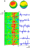
Extraction of the single-trial FRN/FRP and construction of the hemodynamic response functions. A, Example of frontocentral ICs selected for backprojection in one subject. B, Stacked plots of all trials in the same subject as in A ordered by feedback condition; gray line indicates point of grand average maximum difference wave (left column). Parametric hemodynamic response functions extracted from these trials (right column).
Figure 5.
Figure 5.
Result of the conventional, nonparametric GLM analysis. A, F-Map of feedback condition differences for the conventional random-effects analysis with positive, negative, and ambiguous feedback as factor levels. B, F-Map beta estimates (±SEM) for all three conditions in ventral striatal cluster. Whole-brain cluster corrected (11 voxels at p < 0.005); 13 voxels projection depth.
Figure 6.
Figure 6.
Results of the EEG-informed parametric fMRI analyses based on Independent Component Analysis-derived single-trial weights. A, Trial-by-trial coupling of ST-FRN/FRP and BOLD in the ACM: t-map shows significant clusters of activation in MCC, ventral striatum, and temporal gyri. B, Trial-by-trial coupling in the CSM separately for positive, negative, and ambiguous feedback: t-map reveals that coupling between ST-FRN/FRP and BOLD is most pronounced in the positive feedback condition. Whole-brain cluster corrected (11 voxels at p < 0.005); 19 voxels projection depth.

References

    1. Allen PJ, Polizzi G, Krakow K, Fish DR, Lemieux L. Identification of EEG events in the MR scanner: the problem of pulse artifact and a method for its subtraction. Neuroimage. 1998;8:229–239. doi: 10.1006/nimg.1998.0361. - DOI - PubMed
    1. Allen PJ, Josephs O, Turner R. A method for removing imaging artifact from continuous EEG recorded during functional MRI. Neuroimage. 2000;12:230–239. doi: 10.1006/nimg.2000.0599. - DOI - PubMed
    1. Amiez C, Neveu R, Warrot D, Petrides M, Knoblauch K, Procyk E. The location of feedback-related activity in the midcingulate cortex is predicted by local morphology. J Neurosci. 2013;33:2217–2228. doi: 10.1523/JNEUROSCI.2779-12.2013. - DOI - PMC - PubMed
    1. Baker TE, Holroyd CB. Dissociated roles of the anterior cingulate cortex in reward and conflict processing as revealed by the feedback error-related negativity and N200. Biol Psychol. 2011;87:25–34. doi: 10.1016/j.biopsycho.2011.01.010. - DOI - PubMed
    1. Bellebaum C, Koch B, Schwarz M, Daum I. Focal basal ganglia lesions are associated with impairments in reward-based reversal learning. Brain. 2008;131:829–841. doi: 10.1093/brain/awn011. - DOI - PubMed

Publication types

LinkOut - more resources