

Synergistic effects of ischemia and β-amyloid burden on cognitive decline in patients with subcortical vascular mild cognitive impairment
- PMID: 24554306
- PMCID: PMC5849078
- DOI: 10.1001/jamapsychiatry.2013.4506
Synergistic effects of ischemia and β-amyloid burden on cognitive decline in patients with subcortical vascular mild cognitive impairment
Abstract
Importance: Cerebrovascular disease (CVD) and Alzheimer disease are significant causes of cognitive impairment in the elderly. However, few studies have evaluated the relationship between CVD and β-amyloid burden in living humans or their synergistic effects on cognition. Thus, there is a need for better understanding of mild cognitive impairment (MCI) before clinical deterioration begins.
Objective: To determine the synergistic effects of β-amyloid burden and CVD on cognition in patients with subcortical vascular MCI (svMCI).
Design, setting, and participants: A cross-sectional study was conducted using a hospital-based sample at a tertiary referral center. We prospectively recruited 95 patients with svMCI; 67 of these individuals participated in the study. Forty-five patients with amnestic MCI (aMCI) were group matched with those with svMCI by the Clinical Dementia Rating Scale Sum of Boxes.
Main outcomes and measures: We measured β-amyloid burden using positron emission tomography with carbon 11-labeled Pittsburgh Compound B (PiB). Cerebrovascular disease was quantified as white matter hyperintensity volume detected by magnetic resonance imaging fluid-attenuated inversion recovery. Detailed neuropsychological tests were performed to determine the level of patients' cognitive impairment.
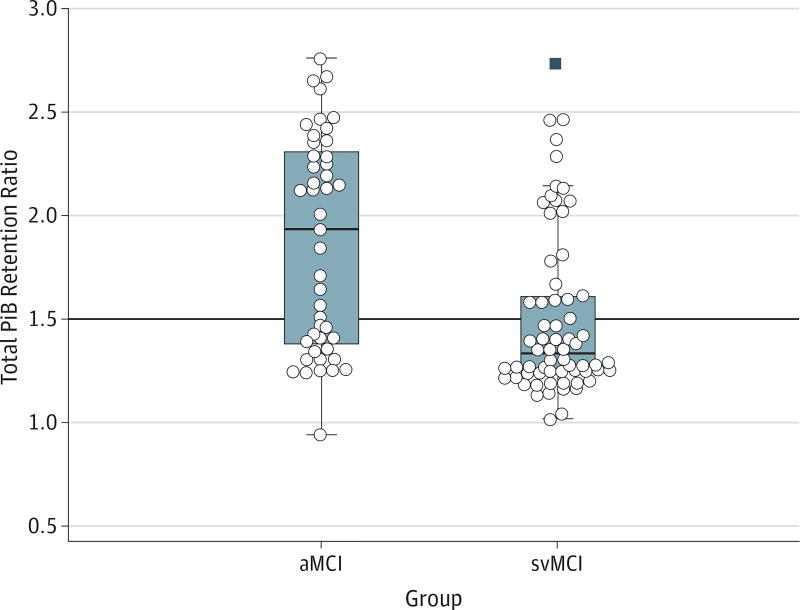
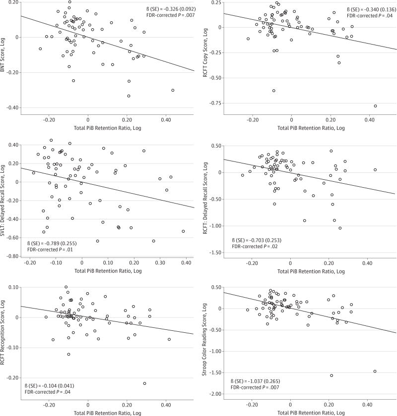
Results: On evaluation, 22 of the svMCI group (33%) and 28 of the aMCI group (62%) were found to be PiB positive. The mean PiB retention ratio was lower in patients with svMCI than in those with aMCI. In svMCI, the PiB retention ratio was associated with cognitive impairments in multiple domains, including language, visuospatial, memory, and frontal executive functions, but was associated only with memory dysfunction in aMCI. A significant interaction between PiB retention ratio and white matter hyperintensity volume was found to affect visuospatial function in patients with svMCI.
Conclusions and relevance: Most patients with svMCI do not exhibit substantial amyloid burden, and CVD does not increase β-amyloid burden as measured by amyloid imaging. However, in patients with svMCI, amyloid burden and white matter hyperintensity act synergistically to impair visuospatial function. Therefore, our findings highlight the need for accurate biomarkers, including neuroimaging tools, for early diagnosis and the need to relate these biomarkers to cognitive measurements for effective use in the clinical setting.
Conflict of interest statement
Figures
References
-
- Frisoni GB, Galluzzi S, Bresciani L, Zanetti O, Geroldi C. Mild cognitive impairment with subcortical vascular features: clinical characteristics and outcome. J Neurol. 2002;249(10):1423–1432. - PubMed
-
- O’Brien JT, Erkinjuntti T, Reisberg B, et al. Vascular cognitive impairment. Lancet Neurol. 2003;2(2):89–98. - PubMed
-
- Wentzel C, Rockwood K, MacKnight C, et al. Progression of impairment in patients with vascular cognitive impairment without dementia. Neurology. 2001;57(4):714–716. - PubMed
-
- Seo SW, Cho SS, Park A, Chin J, Na DL. Subcortical vascular versus amnestic mild cognitive impairment: comparison of cerebral glucose metabolism. J Neuroimaging. 2009;19(3):213–219. - PubMed
-
- Seo SW, Ahn J, Yoon U, et al. Cortical thinning in vascular mild cognitive impairment and vascular dementia of subcortical type. J Neuroimaging. 2010;20(1):37–45. - PubMed
Publication types
MeSH terms
Substances
Grants and funding
LinkOut - more resources
Full Text Sources
Other Literature Sources
Medical
