. 2014 May;88(9):4962-75.
doi: 10.1128/JVI.00063-14. Epub 2014 Feb 19.

Epstein-Barr virus BALF3 has nuclease activity and mediates mature virion production during the lytic cycle

Affiliations

Epstein-Barr virus BALF3 has nuclease activity and mediates mature virion production during the lytic cycle

Shih-Hsin Chiu et al. J Virol. 2014 May.

Abstract

Epstein-Barr virus (EBV) lytic replication involves complex processes, including DNA synthesis, DNA cleavage and packaging, and virion egress. These processes require many different lytic gene products, but the mechanisms of their actions remain unclear, especially for DNA cleavage and packaging. According to sequence homology analysis, EBV BALF3, encoded by the third leftward open reading frame of the BamHI-A fragment in the viral genome, is a homologue of herpes simplex virus type 1 UL28. This gene product is believed to possess the properties of a terminase, such as nucleolytic activity on newly synthesized viral DNA and translocation of unit length viral genomes into procapsids. In order to characterize EBV BALF3, the protein was produced by and purified from recombinant baculoviruses and examined in an enzymatic reaction in vitro, which determined that EBV BALF3 acts as an endonuclease and its activity is modulated by Mg(2+), Mn(2+), and ATP. Moreover, in EBV-positive epithelial cells, BALF3 was expressed and transported from the cytoplasm into the nucleus following induction of the lytic cycle, and gene silencing of BALF3 caused a reduction of DNA packaging and virion release. Interestingly, suppression of BALF3 expression also decreased the efficiency of DNA synthesis. On the basis of these results, we suggest that EBV BALF3 is involved simultaneously in DNA synthesis and packaging and is required for the production of mature virions.

Importance: Virus lytic replication is essential to produce infectious virions, which is responsible for virus survival and spread. This work shows that an uncharacterized gene product of the human herpesvirus Epstein-Barr virus (EBV), BALF3, is expressed during the lytic cycle. In addition, BALF3 mediates an endonucleolytic reaction and is involved in viral DNA synthesis and packaging, leading to influence on the production of mature virions. According to sequence homology and physical properties, the lytic gene product BALF3 is considered a terminase in EBV. These findings identify a novel viral gene with an important role in contributing to a better understanding of the EBV life cycle.


Figures

FIG 1
FIG 1
Sequence alignment of putative conserved motifs in HHV terminase subunits and purification of EBV BALF3. (A) The amino acid (aa) sequences of EBV BALF3 and its homologues encoded by HSV-1, HSV-2, VZV, HCMV, HHV-6, HHV-7, and KSHV were aligned by NPS@ (Network Protein Sequence Analysis; http://npsa-pbil.ibcp.fr). The numbering of amino acid residues is based on the BALF3 sequence, and the identical, strongly similar, and weakly similar amino acid residues are red, green, and blue, respectively. (B) The protein extracts of Sf9 cells were harvested after infection with the wild-type (WT) or recombinant His-BALF3 baculovirus at 28°C for 2 days and subjected to Western blotting with antibodies specific to His. MW, molecular mass; Mock, mock infection. (C) The His-BALF3 protein was purified from the extract of infected Sf9 cells with Ni-NTA resin, and the lysate-resin mixture was washed with 50 mM imidazole and then eluted in the presence of 250 mM imidazole. All of the soluble fractions were collected and subjected to SDS-PAGE with SYPRO Ruby staining. CL, cell lysate; FT, flowthrough; W, wash; E, elution.
FIG 2
FIG 2
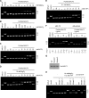
Nuclease activity of EBV BALF3. Purified BALF3 protein was incubated with 0.5 μg pBS-TR and the indicated concentrations of MgCl2 (A), MnCl2 (B), or ATP (C) at 37°C for 1 h prior to agarose gel electrophoresis with EtBr staining. Under the same incubation conditions, the indicated concentrations of ATP were combined with either 10 mM MgCl2 (D) or 0.1 mM MnCl2 (E) to determine the nuclease activity of BALF3. (F) The nuclease activity of the purified BALF3 protein at the indicated doses was assayed in the presence of 10 mM MgCl2 and 12 mM ATP with either pBS-TR or pBlueScript incubation. The intensity of each band was quantified by Gel-Pro Analyzer (Media Cybernetics) and is presented as a percentage. (G) Equal amounts of the purified wild-type (WT), zinc finger motif mutant (mutant 1), and ATP-binding motif mutant (mutant 2) His-BALF3 proteins were identified by Western blotting with antibodies specific to His. (H) The nuclease activity of the purified BALF3 proteins was assayed in the presence of 10 mM MgCl2 and 12 or 20 mM ATP. The open circular, linear, and supercoiled forms of the plasmids are shown as OC, Lin, and SC, respectively. Digestion with HindIII was used as a control for the linear form of the plasmids. ddH2O, double-distilled water.
FIG 3
FIG 3
Expression of EBV BALF3 in EBV-positive cells during the lytic cycle. The mRNA extracts of TW01 and NA cells were harvested at 0, 3, 6, 12, 24, 36, 48, 60, and 72 h after induction with 40 ng/ml TPA and 3 mM SB and subjected to qRT-PCR with primers corresponding to BALF3. The ratios of gene expression levels are shown as fold changes relative to the group of NA cells at 0 h postinduction. Data are presented as means ± standard deviations.
FIG 4
FIG 4
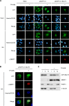
Subcellular localization of EBV BALF3 in EBV-positive cells during the lytic cycle. TW01 and NA cells were transiently transfected with pEGFP-C1-BALF3, followed by induction with 40 ng/ml TPA and 3 mM SB for 36 h, and the images shown were obtained by indirect immunofluorescence staining and photographed using a fluorescence microscope (A) and a confocal microscope (B). The detection of EBV BMRF1 with the specific antibody indicates lytic cycle activation, and the nuclei of the cells were stained with Hoechst 33258. (C) Protein extracts of cells expressing GFP-BALF3 were separated into input (I), cytosolic (C), and nuclear (N) fractions and subjected to Western blotting. After electrophoresis, GFP-BALF3 was detected by an antibody specific to GFP, and PARP1 and α-tubulin were used as markers for the nucleus and the cytoplasm, respectively.
FIG 5
FIG 5
Production of mature virions in EBV-positive cells with BALF3 knockdown. (A) NA cells were transiently transfected with 10 nM Negative Control Duplexes (NC) or the indicated concentrations of siBALF3-1, -2, or -3 prior to treatment with 40 ng/ml TPA and 3 mM SB, and mRNA extracts were harvested at 24 h and subjected to RT-PCR with primers corresponding to BALF3 and other lytic gene products (BZLF1, BRLF1, BALF5, and BMRF1). The amplicons were displayed by agarose gel electrophoresis with EtBr staining. ddH2O, double-distilled water. (B) NA cell supernatants were collected after the transfection of each siRNA at a concentration of 10 nM at 72 h postinduction, and the number of virions was determined by qPCR with BALF5 primers. Data are presented as means ± standard deviations. Student's t test was used to determine the difference between two groups. *, P < 0.01; **, P < 0.001.
FIG 6
FIG 6
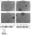
Maturation of capsids in EBV-positive cells with BALF3 knockdown. NA cells were treated with 40 ng/ml TPA and 3 mM SB after the transient transfection of 10 nM Negative Control Duplexes (NC), siBALF3-1, siBALF3-2, or siBALF3-3. (A) At 48 h postinduction, the cells were fixed and embedded in Spurr's resin and the samples were ultrathin sectioned and subjected to transmission electron microscopy. The black arrowheads, white arrows, and black arrows indicate A-, B-, and C-type capsids, respectively. Nuc, nucleus; Cyt, cytoplasm. The scale bar corresponds to 500 nm. (B) At 48 h postinduction, capsids were purified from the cells with NC transfection and separated by sucrose gradient ultracentrifugation, and the fractions were subjected to Western blotting analysis with the antibody specific to EBV VCA. (C and D) Western blotting analysis of fractions obtained by sucrose gradient ultracentrifugation compares the amount of capsids harvested from TPA- and SB-treated cells harboring NC, siBALF3-1, siBALF3-2, or siBALF3-3 to each other. The intensity of each band was quantified by Gel-Pro Analyzer and is presented as a relative fold change.
FIG 7
FIG 7
Examination of viral DNA replication in EBV-positive cells with BALF3 knockdown. (A) NA cells were transiently transfected with 10 nM Negative Control Duplexes (NC), siBALF3-1, siBALF3-2, or siBALF3-3 prior to treatment with 40 ng/ml TPA and 3 mM SB, and mRNA extracts were harvested at 24 and 48 h and subjected to RT-PCR with primers corresponding to BALF3. The amplicons were displayed by agarose gel electrophoresis with EtBr staining. ddH2O, double-distilled water. (B) NA cells were extracted after the transfection of each siRNA at a concentration of 10 nM at 24 and 48 h postinduction, and the EBV genome copy number was measured by qPCR with the BALF5 primers. Data are presented as means ± standard deviations. Student's t test was used to determine the difference between two groups. *, P < 0.01; **, P < 0.001. (C) NA cells were transiently transfected with 10 nM each siRNA, followed by induction. At 48 h postinduction, the cells were extracted and separated in a Gardella gel. After electrophoresis, the gel was subjected to Southern blotting and hybridization with the 32P-radiolabeled random-primed EBV BamHI-W fragment. B95-8, induced with 40 ng/ml TPA and 3 mM SB, and Raji cells were used as the positive controls of linear and episomal EBV DNA, respectively.

References

    1. Nonoyama M, Pagano JS. 1972. Separation of Epstein-Barr virus DNA from large chromosomal DNA in non-virus-producing cells. Nat. New Biol. 238:169–171. 10.1038/newbio238169a0 - DOI - PubMed
    1. Nonoyama M, Tanaka A. 1975. Plasmid DNA as a possible state of Epstein-Barr virus genomes in nonproductive cells. Cold Spring Harbor Symp. Quant. Biol. 39:807–810 - PubMed
    1. Adams A, Lindahl T. 1975. Epstein-Barr virus genomes with properties of circular DNA molecules in carrier cells. Proc. Natl. Acad. Sci. U. S. A. 72:1477–1481. 10.1073/pnas.72.4.1477 - DOI - PMC - PubMed
    1. Yates J, Warren N, Reisman D, Sugden B. 1984. A cis-acting element from the Epstein-Barr viral genome that permits stable replication of recombinant plasmids in latently infected cells. Proc. Natl. Acad. Sci. U. S. A. 81:3806–3810. 10.1073/pnas.81.12.3806 - DOI - PMC - PubMed
    1. Yates JL, Warren N, Sugden B. 1985. Stable replication of plasmids derived from Epstein-Barr virus in various mammalian cells. Nature 313:812–815. 10.1038/313812a0 - DOI - PubMed

Publication types

MeSH terms

LinkOut - more resources