

Fetal ascites and hydrometrocolpos due to persistent urogenital sinus and cloaca: a rare congenital anomaly and review of literature
- PMID: 24554677
- PMCID: PMC3931976
- DOI: 10.1136/bcr-2013-202231
Fetal ascites and hydrometrocolpos due to persistent urogenital sinus and cloaca: a rare congenital anomaly and review of literature
Abstract
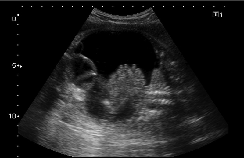
Fetal ascites can occur due to many heterogeneous disorders. Its association with hydrometrocolpos because of persistent urogenital sinus and cloaca is extremely rare. A 29-year-old primigravida presented at 32 weeks of gestation with ultrasonographic evidence of fetal ascites, a cystic pelvic mass, hydronephrosis and oligohydramnios. Fetal ascites in this case was due to fetal urine draining through fallopian tubes into the abdomen as a result of vesicovaginal fistula and distal vaginal atresia. The antenatal ultrasound results along with autopsy findings are discussed. Though rare, a persistent urogenital sinus is to be suspected in isolated fetal ascites cases where the viral tests are negative and there is no evidence of cardiac anomalies as this is a treatable anomaly if diagnosed at early gestational age.
Figures


Similar articles
-
Urinary ascites due to persistent urogenital sinus: A case report and review of literature.J Neonatal Perinatal Med. 2014;7(1):75-9. doi: 10.3233/NPM-1474413. J Neonatal Perinatal Med. 2014. PMID: 24815709 Review.
-
Prenatal ultrasonographic features of persistent urogenital sinus with hydrometrocolpos and ascites.Arch Gynecol Obstet. 2008 Nov;278(5):493-6. doi: 10.1007/s00404-008-0598-3. Epub 2008 Feb 28. Arch Gynecol Obstet. 2008. PMID: 18305949
-
Prenatal diagnosis of persistent urogenital sinus with duplicated hydrometrocolpos and ascites--a case report.Fetal Diagn Ther. 2010;28(4):229-32. doi: 10.1159/000317278. Epub 2010 Aug 18. Fetal Diagn Ther. 2010. PMID: 20720389
-
Prenatal diagnosis of persistent cloaca with hydrometrocolpos and ascites by magnetic resonance imaging in one fetus of a dizygotic twin pregnancy.Taiwan J Obstet Gynecol. 2010 Sep;49(3):385-6. doi: 10.1016/S1028-4559(10)60082-4. Taiwan J Obstet Gynecol. 2010. PMID: 21056332 No abstract available.
-
Isolated hydrometrocolpos and cloacal malformation: can we prenatally distinguish them?-A case report and literature review.Arch Gynecol Obstet. 2025 May;311(5):1467-1474. doi: 10.1007/s00404-025-08004-8. Epub 2025 Apr 2. Arch Gynecol Obstet. 2025. PMID: 40172608 Free PMC article. Review.
Cited by
-
Effectiveness of Prenatal Intervention on the Outcome of Diseases That Have a Postnatal Urological Impact.Front Pediatr. 2019 Apr 2;7:118. doi: 10.3389/fped.2019.00118. eCollection 2019. Front Pediatr. 2019. PMID: 31001504 Free PMC article. Review.
-
An unusual cause of neonatal ascites.BMJ Case Rep. 2017 Jun 2;2017:bcr2017219882. doi: 10.1136/bcr-2017-219882. BMJ Case Rep. 2017. PMID: 28576918 Free PMC article. No abstract available.
-
Successful management of giant hydrocolpos in a limited-resource setting.Oxf Med Case Reports. 2018 Jul 4;2018(7):omy031. doi: 10.1093/omcr/omy031. eCollection 2018 Jul. Oxf Med Case Reports. 2018. PMID: 29992032 Free PMC article.
-
Hydrometrocolpos etiology and management: past beckons the present.Pediatr Surg Int. 2018 Mar;34(3):249-261. doi: 10.1007/s00383-017-4218-9. Epub 2017 Nov 24. Pediatr Surg Int. 2018. PMID: 29177625 Review.
-
Diagnostic imaging and cataloguing of female genital malformations.Insights Imaging. 2016 Oct;7(5):713-26. doi: 10.1007/s13244-016-0515-4. Epub 2016 Aug 9. Insights Imaging. 2016. PMID: 27507534 Free PMC article. Review.
References
-
- Leung WC, Lam YH, Tang MH. Isolated foetal ascitis. Hong Kong Med J 2001;7:432–4 - PubMed
-
- Arena F, Romero C, Cruccetti A, et al. The neonatal management and surgical correction of urinary hydrometrocolpos caused by a persistent urogenital sinus. BJU Int 1999;84:1063–8 - PubMed
-
- Gul A, Yildirim G, Gedikbasi A, et al. Prenatal ultrasonographic features of persistent urogenital sinus with hydrometrocolpos and ascitis. Arch Gynecol Obstet 2008;278:493–6 - PubMed
-
- Blask AR, Sanders RC, Gearhart JP. Obstructed uterovaginal anomalies: demonstration with sonography. Part I. Neonates and infants. Radiology 1991;179:79–83 - PubMed
Publication types
MeSH terms
Supplementary concepts
LinkOut - more resources
Full Text Sources
Other Literature Sources
Medical