. 2014 Apr 4;289(14):9502-18.
doi: 10.1074/jbc.M113.505743. Epub 2014 Feb 19.

Slit diaphragm protein Neph1 and its signaling: a novel therapeutic target for protection of podocytes against glomerular injury

Affiliations

Slit diaphragm protein Neph1 and its signaling: a novel therapeutic target for protection of podocytes against glomerular injury

Ehtesham Arif et al. J Biol Chem. .

Abstract

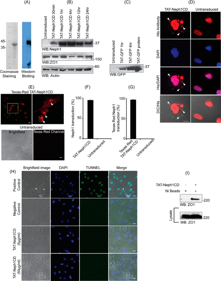
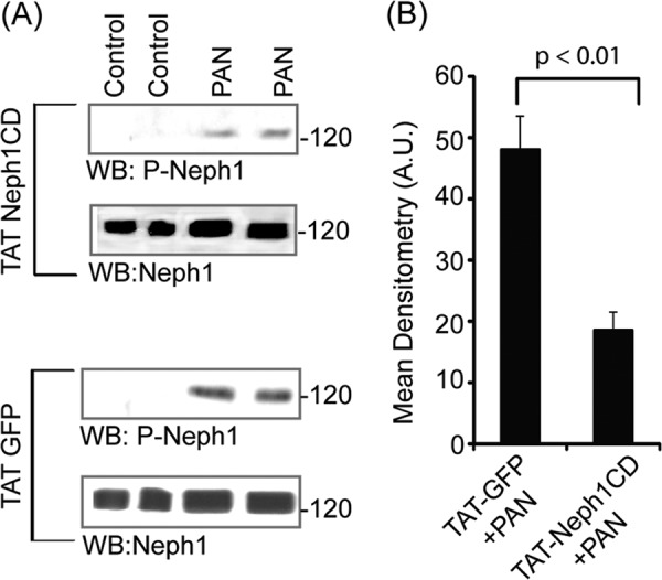
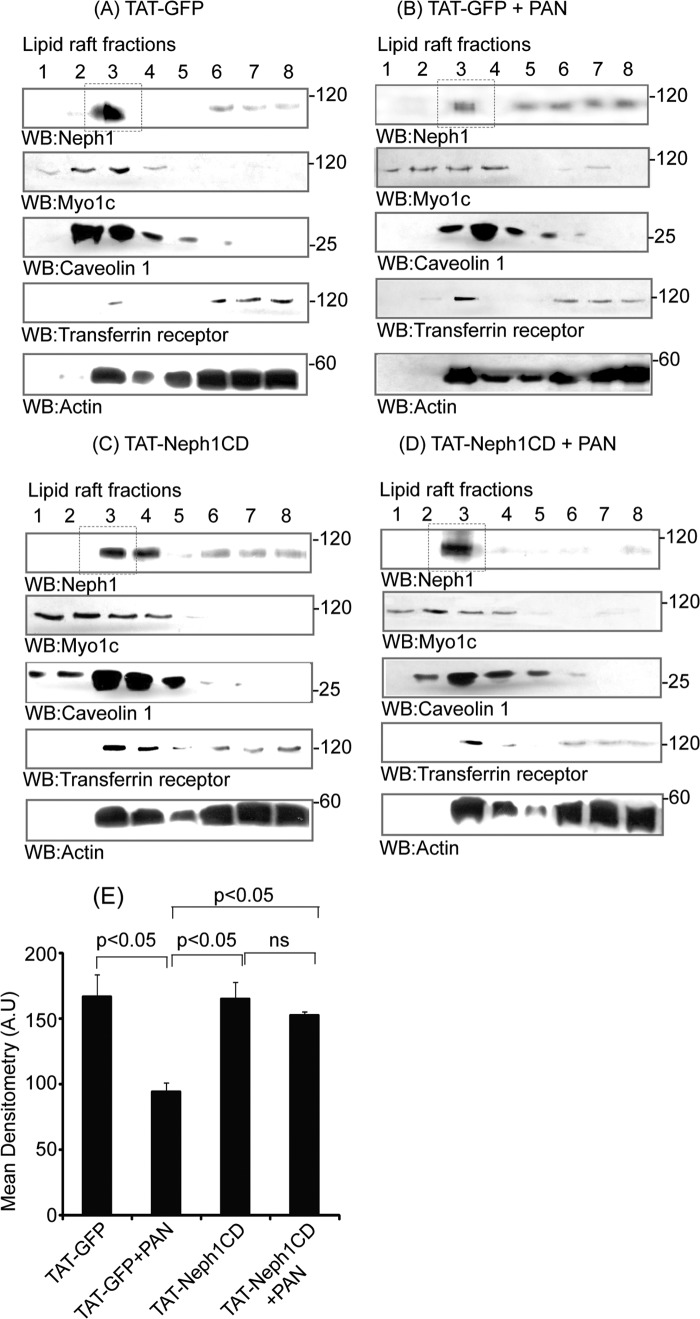
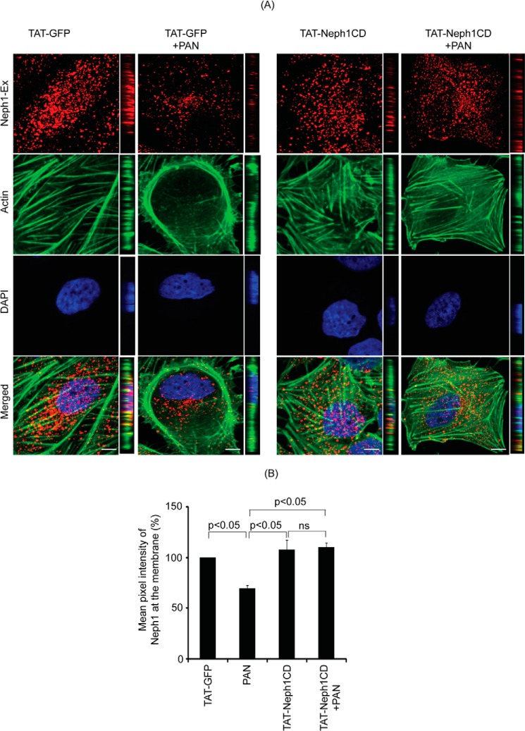
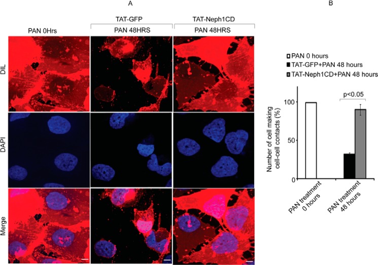
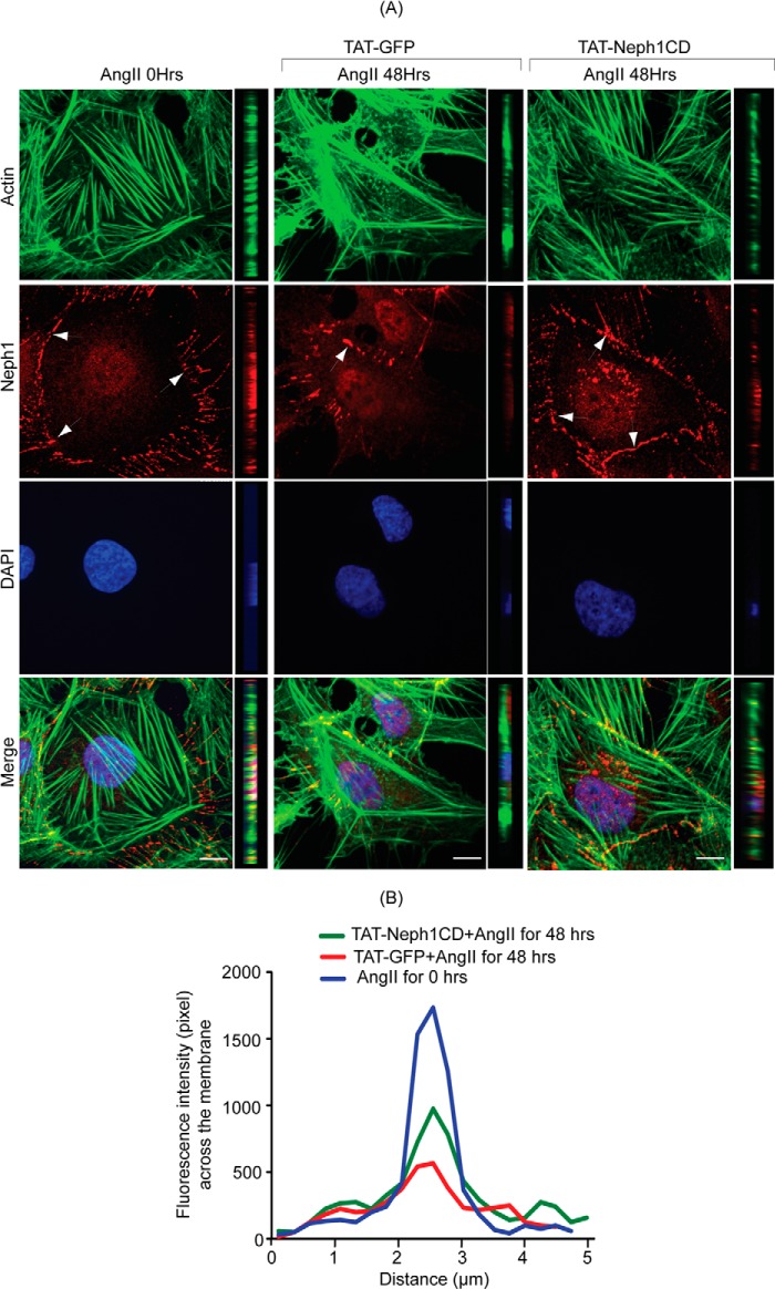
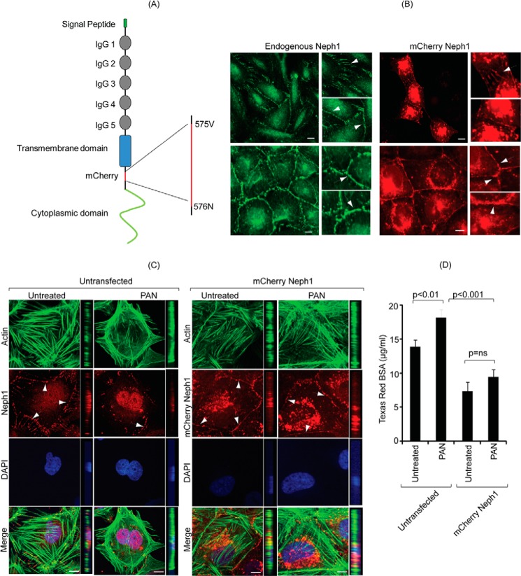
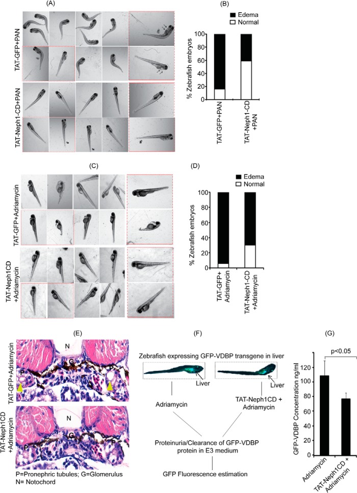
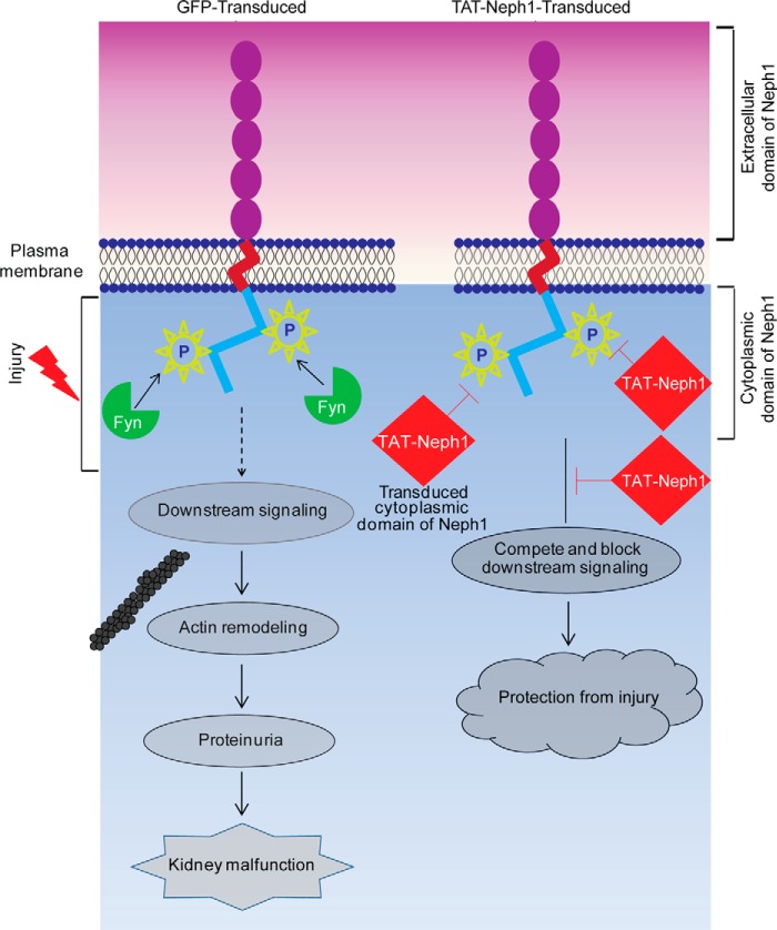
Podocytes are specialized epithelial cells that are critical components of the glomerular filtration barrier, and their dysfunction leads to proteinuria and renal failure. Therefore, preserving podocyte function is therapeutically significant. In this study, we identified Neph1 signaling as a therapeutic target that upon inhibition prevented podocyte damage from a glomerular injury-inducing agent puromycin aminonucleoside (PAN). To specifically inhibit Neph1 signaling, we used a protein transduction approach, where the cytoplasmic domain of Neph1 (Neph1CD) tagged with a protein transduction domain trans-activator of transcription was transduced in cultured podocytes prior to treatment with PAN. The PAN-induced Neph1 phosphorylation was significantly reduced in Neph1CD-transduced cells; in addition, these cells were resistant to PAN-induced cytoskeletal damage. The biochemical analysis using subfractionation studies showed that unlike control cells Neph1 was retained in the lipid raft fractions in the transduced cells following treatment with PAN, indicating that transduction of Neph1CD in podocytes prevented PAN-induced mislocalization of Neph1. In accordance, the immunofluorescence analysis further suggested that Neph1CD-transduced cells had increased ability to retain endogenous Neph1 at the membrane in response to PAN-induced injury. Similar results were obtained when angiotensin was used as an injury-inducing agent. Consistent with these observations, maintaining high levels of Neph1 at the membrane using a podocyte cell line overexpressing chimeric Neph1 increased the ability of podocytes to resist PAN-induced injury and PAN-induced albumin leakage. Using a zebrafish in vivo PAN and adriamycin injury models, we further demonstrated the ability of transduced Neph1CD to preserve glomerular function. Collectively, these results support the conclusion that inhibiting Neph1 signaling is therapeutically significant in preventing podocyte damage from glomerular injury.

Keywords: Actin; Cell Signaling; Kidney; Neph1; Phosphorylation; Podocytes; Renal Injury.


Figures

FIGURE 1.
FIGURE 1.
TAT-Neph1-CD is transduced in podocytes and remains functional. A, recombinantly expressed TAT-Neph1CD was purified, and its purity and identity were assessed by staining with Coomassie and Western blotting with Neph1 antibody, respectively. B, TAT-Neph1CD protein (5 μg/ml) was transduced in cultured podocytes for the indicated time periods ranging from 30 min to 24 h and subjected to Western blotting (WB) with Neph1 antibody. The protein rapidly transduced inside the cells within 30 min and was detected even at 24 h post-transduction. C, in a similar fashion, TAT-GFP was transduced for indicated times and was used as a control. D, TAT-Neph1CD was transduced in cultured human podocytes for a period of 30 min, washed, and fixed with 4% PFA (1× PBS). His primary antibody and Alexa Fluor 594 secondary antibodies were used to label the transduced TAT-Neph1CD (arrows), and the immunofluorescence imaging analysis was performed using a fluorescent microscope (×60 magnification). Additionally, the cells were visualized by differential interference contrast (DIC) microscopy. E, TAT-Neph1CD protein was labeled with Texas red dye and transduced in cultured human podocytes for 30 min. The cells were then washed and fixed with 4% PFA (in 1× PBS) and analyzed by immunofluorescence imaging that showed significant accumulation of fluorescent TAT-Neph1CD inside the transduced podocytes as compared with the untransduced control podocytes. F, statistical analysis of the TAT-Neph1CD transduction from D suggested that 94.6 ± 1.5% of the podocytes were transduced with this protein. G, statistical analysis of the Texas red TAT-Neph1CD transduction suggested that 96.3 ± 1.5% of podocytes were transduced with this protein. H, TUNEL assay was performed on transduced and untransduced podocytes. No apoptosis could be seen in the podocytes that were transduced with 10 times (50 μg/ml) higher than the usual dose (5 μg/ml) of TAT-Neph1CD in podocytes. Bright field and DAPI images were also obtained to visualize the overall cell population. I, ability of transduced TAT-Neph1CD to interact with endogenous ZO-1 was examined in a pulldown experiment using Ni-NTA beads. Podocytes transduced with TAT-Neph1CD were lysed, and the Neph1-ZO-1 complex was pulled down using nickel beads, separated on SDS-PAGE, and analyzed by Western blotting to detect the presence of ZO-1 in the complex. Scale bars, 10 μm (D), 20 μm (E); 20 μm (H).
FIGURE 2.
FIGURE 2.
Transduced TAT-Neph1CD inhibited tyrosine phosphorylation of endogenous Neph1. A, podocytes transduced with either TAT-GFP or TAT-Neph1CD were treated with PAN for a period of 48 h, and the cell lysates were analyzed for the presence of phosphorylated Neph1 by Western blotting (WB) with phospho-Neph1 (p-Neph1) and Neph1 antibodies. The transduction of TAT-Neph1CD in podocytes significantly reduced PAN-induced endogenous Neph1 phosphorylation as compared with the control. B, further densitometric analysis revealed a 60–70% decrease of the PAN-induced endogenous Neph1 phosphorylation in cells transduced with TAT-Neph1CD as compared with the TAT-GFP-transduced cells.
FIGURE 3.
FIGURE 3.
Redistribution of endogenous Neph1 to non-lipid raft fractions in response to PAN was inhibited by TAT-Neph1CD transduction. A–D, TAT-GFP- and TAT-Neph1CD-transduced podocytes were treated with or without PAN and subjected to subfractionation using Optiprep gradient. The lysates were run on an SDS-PAGE, transferred to nitrocellulose, and Western blotted (WB) using Neph1, Myo1c, caveolin 1, transferrin receptor, and actin antibodies. PAN-induced redistribution of endogenous Neph1 to non-lipid raft fractions (5–8) is shown in B, whereas treatment with TAT-Neph1CD (D) retained Neph1 in the lipid raft fraction (3). E, densitometric analysis of PAN-treated cells suggested a 70% loss of Neph1 from the lipid rafts (marked in dotted box), which was restored by the transduction of TAT-Neph1CD.
FIGURE 4.
FIGURE 4.
Transduction of TAT-Neph1CD prevents PAN-induced loss of Neph1 from podocyte cell membrane. A, transduced cells were analyzed by immunofluorescence using antibodies directed against actin and the extracellular domain of Neph1 (Neph1-Ex). Confocal images were collected at ×60 magnification and presented after deconvolution in XY and XZ orientation. B, analysis of mean pixel intensity at the membrane of transduced cells suggests that PAN treatment significantly reduced Neph1 localization at the membrane (p < 0.05) in control cells (TAT-GFP+PAN), whereas the cells transduced with TAT-Neph1CD maintained a robust localization of Neph1 at the podocyte cell membrane and were unaffected by treatment with PAN. ns, nonsignificant. Scale bar, A, 10 μm.
FIGURE 5.
FIGURE 5.
TAT-Neph1CD-transduced podocytes are resistant to PAN-induced cytoskeletal damage. A and B, TAT-GFP- and TAT-Neph1CD-transduced podocytes were treated with PAN for indicated time periods (0–48 h) and immunostained with phalloidin (Alexa 488) and Neph1 (Alexa 594), and mounted with DAPI. Confocal imaging showed that PAN treatment resulted in drastic changes to the actin cytoskeleton in TAT-GFP-transduced cells. Additionally, the distribution of junctional proteins, including Neph1, was significantly altered in these cells. In contrast, the PAN treatment of podocytes transduced with TAT-Neph1CD showed minimal alteration to the actin cytoskeleton with increased localization of Neph1 at the junctions. Confocal images were collected at ×60 magnification and presented after deconvolution in XY and XZ orientation. B, pixel intensity profile was created by placing a line spanning the cell-cell junction and analyzed using ImageJ software. This analysis suggested that the intensity of Neph1 at the cell-cell junctions was maintained in TAT-Neph1CD-transduced cells treated with PAN (p < 0.05). Scale bar, 10 μm (A).
FIGURE 6.
FIGURE 6.
Cell-cell contacts were preserved in TAT-Neph1CD-transduced podocytes treated with PAN. A and B, podocytes were labeled with Vybrantô DiI cell membrane labeling dye for easier visualization of cell boundaries. Number of podocyte cells that were engaged in cell-cell contacts were identified and analyzed. The cell-cell contacts were calculated by analyzing 100 cells in each group (in triplicate), and the cells with more than 50% of their surface (defined manually using actin and membrane DiI staining images) in contact with the neighboring cells were considered positive. The podocytes transduced with TAT-GFP showed significant reduction in the cell-cell contacts, and thus the number of cells involved in contact formation were significantly reduced in response to 48 h of PAN treatment; in contrast, the cell-cell contacts were well preserved in the TAT-Neph1CD-transduced cells treated with PAN. Scale bar, 10 μm (A).
FIGURE 7.
FIGURE 7.
TAT-Neph1CD-transduced podocytes are resistant to cytoskeletal damage induced by Ang II. A and B, TAT-GFP- and TAT-Neph1CD-transduced podocytes were treated with Ang II for the indicated time periods (0–48 h) and immunostained with phalloidin (Alexa 488) and Neph1 (Alexa 594) and mounted with DAPI containing mounting media. Confocal imaging revealed that Ang II treatment resulted in changes to the actin cytoskeleton (where increased stress fibers were noted) along with loss of Neph1 at the cell-cell junctions (A). Notably, the transduction of TAT-Neph1CD prevented these changes (see Fig. 5A). Confocal images were collected at ×60 magnification and constructed after deconvolution in XY and XZ orientation. B, quantitation using pixel intensity profile further demonstrated that similar to PAN, the loss of Neph1 at the cell-cell junctions was minimal in TAT-Neph1CD transduced cells treated with Ang II when compared with the TAT-GFP transduced cells treated with Ang II (p < 0.05). Scale bar, 10 μm (A).
FIGURE 8.
FIGURE 8.
Podocytes overexpressing Neph1 are resistant to PAN-induced injury. A, schematic representation of the construction of Cherry-Neph1 construct (where Cherry was introduced after the transmembrane domain and before any potential tyrosine phosphorylation sites in Neph1) is shown. B, expression pattern of Cherry-Neph1 was similar to endogenous Neph1 in podocytes, where both proteins localized at the cell-cell junctions (arrows). C, podocytes without and with stable expression of Cherry-Neph1 were created and subjected to PAN treatment for 48 h. Unlike the control cells transfected with empty vector, the Cherry-Neph1-transfected cells were resistant to PAN-induced cytoskeletal damage, and their cell-cell junctions were well maintained. D, BSA permeability assay was performed with vector-transfected (control) and Cherry-Neph1-overexpressing podocytes that were cultured as a monolayer on Transwell filters and treated with PAN. The passage of albumin across the podocytes monolayer was assessed by a paracellular albumin flux assay using Texas red-labeled albumin. The Cherry-Neph1-overexpressing cells showed increased resistance to PAN-induced albumin leakage as compared with control podocytes. ns, nonsignificant. Scale bar, 20 μm (B); 10 μm (C).
FIGURE 9.
FIGURE 9.
Zebrafish injected with TAT-Neph1CD were resistant to PAN- and Ang II-induced injury. A, PAN (25 mg/ml) was co-injected with 0.25 mg/ml of either TAT-GFP (control) or TAT-Neph1CD (test) in 72-hpf zebrafish via the common cardinal vein, and the development of pericardial edema was monitored at 96 hpf. Significant increase in pericardial edema and curved body shape was noted in the control, whereas the test zebrafish had minimal or no edema, and their body shapes were grossly normal. B, quantitative analysis from three independent experiments suggested about 40% reduction of PAN-induced edema in test zebrafish as compared with the control. C and D, embryos either injected with 0.25 mg/ml of either TAT-GFP (control) or TAT-Neph1CD (test) were grown in the E3 medium containing adriamycin (30.3 mg/liter). The phenotype (pericardial edema an indication of renal abnormality) was observed at 96 hpf (more than 90% embryos develop edema in this model). However, the number of zebrafish embryos with pericardial edema was significantly reduced with TAT-Neph1CD injection. D, quantitative analysis further suggested a 25% decrease in pericardial edema in embryos injected with TAT-Neph1CD when compared with the embryos injected with TAT-GFP (p < 0.05). E, histological analysis of sections from 72 hpf of TAT-GFP-injected embryos treated with adriamycin and stained with hematoxylin and eosin showed prominent dilation of the pronephric tubules, whereas this phenotype was absent in TAT-Neph1CD-injected embryos treated with adriamycin. F, schematic representation of the in vivo filtration assay using transgenic zebrafish expressing GFP-VDBP from liver. Zebrafish injected with TAT-Neph1CD and uninjected were treated with adriamycin to induce glomerular injury, and the excreted GFP-VDBP was estimated in the medium. G, zebrafish embryos injected with TAT-Neph1CD showed a measurable reduction (∼30% with a p value of <0.05) in the excreted GFP-VDBP in the medium when compared with the control embryos.
FIGURE 10.
FIGURE 10.
Schematic presentation of the proposed TAT-Neph1CD action. The tyrosine phosphorylation of endogenous Neph1 is increased in response to PAN that induces downstream signaling leading to cytoskeleton damage and kidney malfunction. Transduction of TAT-Neph1CD competes for endogenous Neph1 interactions, thereby blocking Neph1 phosphorylation and inhibiting the downstream signaling. Thus, the transduced cells are unable to respond to injury and are therefore protected from the PAN-induced podocyte damage.

References

    1. Haraldsson B., Nyström J., Deen W. M. (2008) Properties of the glomerular barrier and mechanisms of proteinuria. Physiol. Rev. 88, 451–487 - PubMed
    1. Pavenstädt H., Kriz W., Kretzler M. (2003) Cell biology of the glomerular podocyte. Physiol. Rev. 83, 253–307 - PubMed
    1. Tryggvason K., Patrakka J., Wartiovaara J. (2006) Hereditary proteinuria syndromes and mechanisms of proteinuria. N. Engl. J. Med. 354, 1387–1401 - PubMed
    1. Tryggvason K., Pikkarainen T., Patrakka J. (2006) Nck links nephrin to actin in kidney podocytes. Cell 125, 221–224 - PubMed
    1. Verma R., Kovari I., Soofi A., Nihalani D., Patrie K., Holzman L. B. (2006) Nephrin ectodomain engagement results in Src kinase activation, nephrin phosphorylation, Nck recruitment, and actin polymerization. J. Clin. Invest. 116, 1346–1359 - PMC - PubMed

Publication types

MeSH terms