. 2014 Mar 15;192(6):2837-45.
doi: 10.4049/jimmunol.1302484. Epub 2014 Feb 19.

Cholesterol crystals induce complement-dependent inflammasome activation and cytokine release

Affiliations

Cholesterol crystals induce complement-dependent inflammasome activation and cytokine release

Eivind O Samstad et al. J Immunol. .

Abstract

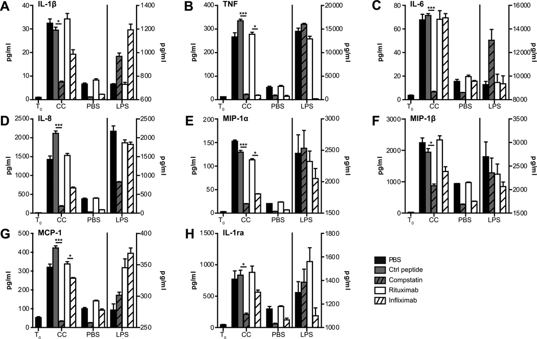
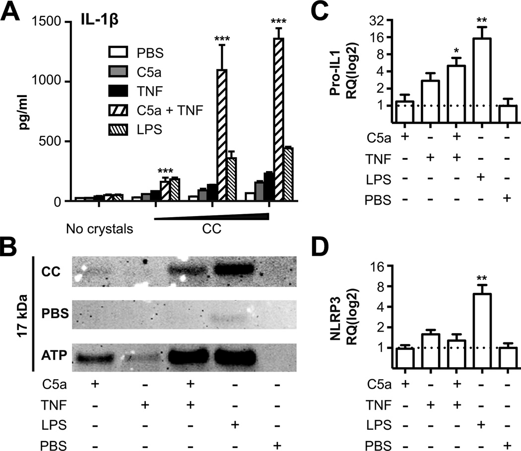
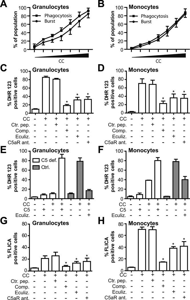
Inflammation is associated with development of atherosclerosis, and cholesterol crystals (CC) have long been recognized as a hallmark of atherosclerotic lesions. CC appear early in the atheroma development and trigger inflammation by NLRP3 inflammasome activation. In this study we hypothesized whether CC employ the complement system to activate inflammasome/caspase-1, leading to release of mature IL-1β, and whether complement activation regulates CC-induced cytokine production. In this study we describe that CC activated both the classical and alternative complement pathways, and C1q was found to be crucial for the activation. CC employed C5a in the release of a number of cytokines in whole blood, including IL-1β and TNF. CC induced minimal amounts of cytokines in C5-deficient whole blood, until reconstituted with C5. Furthermore, C5a and TNF in combination acted as a potent primer for CC-induced IL-1β release by increasing IL-1β transcripts. CC-induced complement activation resulted in upregulation of complement receptor 3 (CD11b/CD18), leading to phagocytosis of CC. Also, CC mounted a complement-dependent production of reactive oxygen species and active caspase-1. We conclude that CC employ the complement system to induce cytokines and activate the inflammasome/caspase-1 by regulating several cellular responses in human monocytes. In light of this, complement inhibition might be an interesting therapeutic approach for treatment of atherosclerosis.


Conflict of interest statement

Disclosures:

Conflict of interest: The co-author, Prof. J.D.L., is the holder of several patent applications on complement inhibitors and the founder of Amyndas Pharmaceuticals, which is developing complement inhibitors for clinical applications. The other authors report no conflict.

Figures

Figure 1
Figure 1. CC activate the alternative- and classical complement pathways
Human serum was incubated at 37°C, for indicated times, in the presence of CC (3 × 10⁷ particles/ml), zymosan and heat aggregated IgG (Zym-IgG) or PBS. (A) The end product in complement activation, TCC, showed a significant increase (***, p < 0.001) in response to CC compared to PBS at every time points, (B) the activation product C3bc, from the common complement component C3 for all three initial pathways, showed a significant increase (***, p < 0.001) at all time points in response to CC compared to PBS, (C) the alternative pathway convertase, C3bBbP, showed a significant increase (***, p < 0.001) in response to CC compared to PBS at every time points. The increase in (D) the common activation product for the classical and lectin pathways, C4bc, and (E) the activation product for the classical pathway, C1rs-C1-INH, did not reach statistical significance. Data plotted are mean ± SEM from six independent experiments with serum from healthy donors. (F) C1q binding to CC, when incubated in plasma for 30 minutes, measured by flow cytometry. (G) TCC in C1q depleted serum (C1q dep.) with or without reconstitution (C1q rec.) (*, p < 0.05) with purified C1q upon CC (1.5 × 10⁷ particles/ml) stimulation, measured by ELISA. One out of six independently performed experiments is shown. In the lower panel, human whole blood was incubated with CC (1.5 × 10⁷ particles/ml) for 30 minutes. (H) Binding of TCC to the crystals was detected using anti C5b-9 and anti-mouse IgG conjugated with Alexa-488, and for C3bc (I) anti-C3bc antibody directly conjugated to FITC. (J) Control IgG2a conjugated to FITC. Scale bars represent 10 µm. Data are representative of two independent experiments. AU = arbitrary units. DIC = Differential Interference Contrast.
Figure 2
Figure 2. CC induce complement-dependent cytokine release
Human whole blood was incubated with CC (3 × 10⁷ particles/ml), PBS or LPS for 6 hours after preincubation with PBS, control peptide, compstatin, anti-TNF infliximab, or control anti-CD20 rituximab at 37°C. Cytokines and chemokines were quantified in plasma by multiplex analysis. T0 represents the start of the experiment. Dataset on the left of the dividing line (T0, CC, PBS) are plotted on the left y-axis, and dataset on the right of the dividing line (LPS) are plotted on the right y-axis. Data plotted are mean ± SD from triplicate determinations in one out of at least six independently performed experiments from healthy donors (*, p < 0.05, ***, p < 0.001).
Figure 3
Figure 3. CC-induced release of cytokines from PBMC and granulocytes
Human PBMC and granulocytes were isolated from whole blood and resuspended in plasma/PBS before incubation with CC (3 × 10⁷ particles/ml) for 5.5 hours. CC-induced production of the cytokines (A) IL-1β, (B) TNF, (C) IL-6, (D) IL-8, (E) MIP-1α, and (F) MCP-1 from PBMC and granulocytes were quantified by multiplex analysis and compared (*, p <0.05). (G) Myeloperoxidase (MPO) from PBMC and granulocytes was detected by ELISA (*, p <0.05). Data plotted are mean ± SD in triplicate determinations in one out of six independent experiments.
Figure 4
Figure 4. CC-induced CD11b expression and cytokine production in whole blood from a C5-deficient person
Whole blood from a C5 deficient donor, or C5 deficient donor reconstituted with purified C5 (50 µg/ml), or blood from three healthy donors were incubated with CC (3 × 10⁷ particles/ml), PBS/HSA or LPS at 37°C. (A) TCC detected by ELISA in plasma after 30 minutes. (B, C) Median fluorescent intensity (MFI) of CD11b on granulocytes and monocytes measured after 15 minutes. Plasma collected after 6 hours incubation was analyzed by multiplex for (D) IL-1β, (E) TNF, (F) IL-6, and (G) IL-8. Mean of triplicate determinations are shown for the C5 defect donor samples, and mean of three control donors are shown. Error bars: ±SD.
Figure 5
Figure 5. Combining C5a and TNF prime PBMC and monocytes for CC-induced IL-1β
Human (A) PBMCs and (B-D) monocytes were primed for 2 hours in 10% heat inactivated human serum with PBS/HSA, C5a, TNF, a combination of the two, or a priming dose of LPS (100 pg/ml) prior to stimulation with CC. (A) IL-1β was detected by ELISA in supernatants 16 hours after stimulation with increasing concentrations of CC (0.15 × 1070.75 × 1071.5 × 107 particles/ml). Combination of C5a and TNF are compared to TNF alone for all concentrations (***, p < 0.001). (B) Cleaved IL-1β detected by western blot in monocytes, primed as indicated by matrix below blot, and stimulated for 6 hours with CC (1.5 × 10⁷ particles/ml), ATP (3 mM) or PBS/HSA. (C) Pro-IL-1β - or (D) NLRP3 mRNA were measured by qRT-PCR and normalized against PBS control (*, p < 0.05, **, p < 0.01). One experiment of at least three performed is shown.
Figure 6
Figure 6. CC-induced up-regulation of CD11b (CR3) and phagocytosis of CC are complement-mediated
Whole blood pre-incubated with the complement inhibitors compstatin (Comp.), eculizumab (Eculiz.), C5a receptor antagonist (C5aR ant.), control peptide (Ctrl. Pep.) or anti-C7 (C7 antibody) was incubated with CC (3 × 10⁷ particles/ml), or PBS/HSA. (A, B) Median fluorescent intensity (MFI) of CD11b on granulocytes and monocytes measured after 15 minutes. (C, D) Phagocytosis was determined based on shift in SSC induced by CC ingestion, as described in Materials and Methods. The decrease compared to the control peptide group in each panel (A-D) was significant (*, p < 0.05) for all comparisons, except for C7 antibody. Results are mean ± SEM, n=6. (E, F) Phagocytosis of CC in granulocytes and monocytes in whole blood from a C5 deficient donor (C5 def.), or C5 deficient donor reconstituted with purified C5 (50 µg/ml). Blood from three controls (Ctrl.) were analyzed in parallel. Results from the C5 deficient donor represent mean ± SD of two experiments performed at two time points.
Figure 7
Figure 7. CC-induced ROS production and caspase-1 activation are complement-mediated
Whole blood was incubated with increasing concentrations of CC (0 – 6 × 10⁷ particles/ml). A dose-dependent relationship between CC and phagocytosis/oxidative burst in granulocytes (A) and monocytes (B) was measured. Whole blood pre-incubated with the complement inhibitors compstatin (Comp.), eculizumab (Eculiz.), C5a receptor antagonist (C5aR ant.), control peptide (Ctrl. Pep.) and incubated with CC (3 × 10⁷ particles/ml) or PBS/HSA. ROS production is shown as % dihydrorhodamine (DHR) 123 positive (C) granulocytes or (D) monocytes. (E, F) CC-induced ROS production in granulocytes and monocytes from a C5 deficient donor, or C5 deficient donor reconstituted with purified C5. Results from the C5 deficient donor represent mean ± SD of two experiments performed at two time points and blood from three controls (Ctrl.) were analyzed in parallel. Activation of caspase-1 was detected as % FLICA positive (G) granulocytes or (H) monocytes in whole blood (mean ± SEM, n=3, *, p < 0.05). The decrease compared to the control peptide group in (C, D) and (G, H) was significant (mean ± SEM, n=6,*, p < 0.05) for all comparisons.

References

    1. Small DM. George Lyman Duff memorial lecture. Progression and regression of atherosclerotic lesions. Insights from lipid physical biochemistry. Arteriosclerosis. 1988;8:103–129. - PubMed
    1. Abela GS. Cholesterol crystals piercing the arterial plaque and intima trigger local and systemic inflammation. J. Clin. Lipidol. 2010;4:156–164. - PubMed
    1. Stary HC, Chandler AB, Dinsmore RE, Fuster V, Glagov S, Insull W, Jr, Rosenfeld ME, Schwartz CJ, Wagner WD, Wissler RW. A definition of advanced types of atherosclerotic lesions and a histological classification of atherosclerosis. A report from the Committee on Vascular Lesions of the Council on Arteriosclerosis, American Heart Association. Arterioscler. Thromb. Vasc. Biol. 1995;15:1512–1531. - PubMed
    1. Duewell P, Kono H, Rayner KJ, Sirois CM, Vladimer G, Bauernfeind FG, Abela GS, Franchi L, Nunez G, Schnurr M, Espevik T, Lien E, Fitzgerald KA, Rock KL, Moore KJ, Wright SD, Hornung V, Latz E. NLRP3 inflammasomes are required for atherogenesis and activated by cholesterol crystals. Nature. 2010;464:1357–1361. - PMC - PubMed
    1. Sheedy FJ, Grebe A, Rayner KJ, Kalantari P, Ramkhelawon B, Carpenter SB, Becker CE, Ediriweera HN, Mullick AE, Golenbock DT, Stuart LM, Latz E, Fitzgerald KA, Moore KJ. CD36 coordinates NLRP3 inflammasome activation by facilitating intracellular nucleation of soluble ligands into particulate ligands in sterile inflammation. Nat. Immunol. 2013;14:812–820. - PMC - PubMed

Publication types

MeSH terms