. 2014 May;111(10):1927-39.
doi: 10.1152/jn.00631.2013. Epub 2014 Feb 19.

In vivo and in vitro analyses of amygdalar function reveal a role for copper

Affiliations

In vivo and in vitro analyses of amygdalar function reveal a role for copper

E D Gaier et al. J Neurophysiol. 2014 May.

Abstract

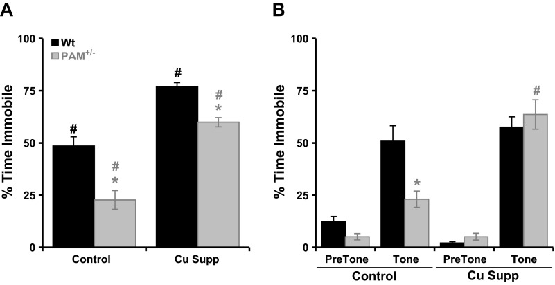
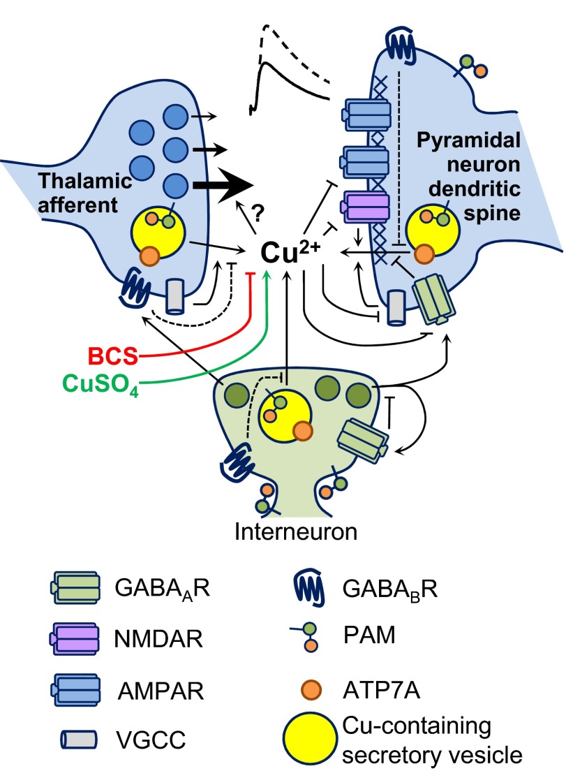
Mice with a single copy of the peptide amidating monooxygenase (Pam) gene (PAM(+/-)) are impaired in contextual and cued fear conditioning. These abnormalities coincide with deficient long-term potentiation (LTP) at excitatory thalamic afferent synapses onto pyramidal neurons in the lateral amygdala. Slice recordings from PAM(+/-) mice identified an increase in GABAergic tone (Gaier ED, Rodriguiz RM, Ma XM, Sivaramakrishnan S, Bousquet-Moore D, Wetsel WC, Eipper BA, Mains RE. J Neurosci 30: 13656-13669, 2010). Biochemical data indicate a tissue-specific deficit in Cu content in the amygdala; amygdalar expression of Atox-1 and Atp7a, essential for transport of Cu into the secretory pathway, is reduced in PAM(+/-) mice. When PAM(+/-) mice were fed a diet supplemented with Cu, the impairments in fear conditioning were reversed, and LTP was normalized in amygdala slice recordings. A role for endogenous Cu in amygdalar LTP was established by the inhibitory effect of a brief incubation of wild-type slices with bathocuproine disulfonate, a highly selective, cell-impermeant Cu chelator. Interestingly, bath-applied CuSO₄ had no effect on excitatory currents but reversibly potentiated the disynaptic inhibitory current. Bath-applied CuSO₄ was sufficient to potentiate wild-type amygdala afferent synapses. The ability of dietary Cu to affect signaling in pathways that govern fear-based behaviors supports an essential physiological role for Cu in amygdalar function at both the synaptic and behavioral levels. This work is relevant to neurological and psychiatric disorders in which disturbed Cu homeostasis could contribute to altered synaptic transmission, including Wilson's, Menkes, Alzheimer's, and prion-related diseases.

Keywords: GABA; fear; learning and memory; peptidylglycine alpha-amidating monooxygenase; synaptic plasticity.


Figures

Fig. 1.
Fig. 1.
Reduced Cu in PAM+/− amygdala. Bilateral amygdalae (Amyg) and dorsal hippocampi (Hipp) were isolated from individual wild-type (Wt) and PAM+/− mice by tissue punch. Tissue samples were analyzed for Cu concentration using inductively coupled plasma mass spectrometry; Cu levels were normalized to protein concentration for comparisons by genotype and brain region. Cu concentrations according to protein are plotted by genotype for amygdala and hippocampus. *P < 0.05, compared with Wt mice within brain region; ‡P < 0.05, compared with amygdala within genotype; n = 7–9 mice/genotype/brain region.
Fig. 2.
Fig. 2.
Cu supplementation does not affect contextual fear conditioning while rescuing PAM+/− responses in cued testing. Two cohorts of Wt and PAM+/− littermates were given either control water or Cu-supplemented water (Cu Supp) for 14 days before conditioning. A: for 1 cohort, percentage of time freezing during the 5-min contextual test was assessed 24 h after conditioning (each minute averaged). B: for the other cohort, percentage of time freezing in the 2 min before (PreTone) and the 3 min during presentation (Tone) of the single 30-s, 72-dB tone (CS) in cued testing was assessed 24 h after conditioning. *P < 0.05, compared with Wt mice within the Cu condition; #P < 0.05, compared with Cu condition within genotype; n = 9–11 mice/genotype/Cu condition.
Fig. 3.
Fig. 3.
Cu supplementation rescues the PAM+/− amygdalar synaptic plasticity deficit. A, left: schematic of a coronal brain slice containing the amygdala; recording electrode (rec) placement in the lateral nucleus of the amygdala (L) and stimulating electrode (stim) placement in thalamic afferent fibers are illustrated. Also depicted for reference: cerebral cortex (Cort), hippocampus (H), thalamus (Th), hypothalamus (Hy), and basolateral and central nuclei of the amygdala (BL and C). Right: synaptic schematic illustrating recording from a lateral amygdala pyramidal neuron (PN), stimulation of thalamic afferents, and feedforward inhibition, which is mediated by GABAergic interneurons (IN) and was blocked in these experiments by inclusion of 100 μM picrotoxin (PTX). B: Wt and PAM+/− littermates were fed control water before amygdalar slice preparation for long-term potentiation (LTP) experiments using an action potential pairing induction paradigm starting at minute 0 (arrow). The time course (1-min bins) of averaged LTP experiments for Wt (black) and PAM+/− (gray) neurons are shown. Normalized rise slopes of the excitatory postsynaptic potentials (NL EPSP slope) were used to measure synaptic efficacy. Baseline values correspond to responses during minutes −5 to 0, and postinduction LTP values correspond to responses during minutes 3040. Traces above plots depict averaged EPSPs corresponding to the time periods outlined by solid (baseline) and dashed (postinduction) lines. C: paired-pulse ratios (PPRs; rise slope ratio EPSP2 to EPSP1) were recorded throughout the LTP time course in B and are plotted in 5-min bins by genotype. Absolute PPR values averaged over the baseline (BL; −5 to 0 min) and postinduction (LTP; 30–40 min) are plotted by genotype in the inset. D and E: current-voltage plots generated from amygdalar neurons from Wt and PAM+/− littermates fed normal (C) and Cu-supplemented (D) water. F: representative voltage responses to the paired LTP induction protocol recorded in amygdalar neurons from Wt mice fed control water vs. Cu-supplemented water. G: Wt and PAM+/− littermates were fed Cu-supplemented water before amygdalar slice preparation for LTP experiments, which were conducted as in B. +P < 0.05, compared with baseline within genotype and Cu condition; NS, not significant compared with baseline (Wilcoxon signed-rank test); *P < 0.05, compared with Wt mice within Cu condition; #P < 0.05, compared with control water within genotype (unpaired t-test); n = 7–8 mice/genotype/Cu condition.
Fig. 4.
Fig. 4.
Bath-applied CuSO4 transiently enhances inhibitory synaptic transmission and restores PAM+/− LTP. A: pyramidal neurons in the lateral amygdala of Wt mice were patched with pipettes filled with intracellular solution containing open voltage-gated Na+ channel blocker QX-314 and voltage-clamped at holding potential (Vh) = −35 mV. Left: synaptic schematic depicting the recording paradigm. Current responses to thalamic afferent stimulation were recorded for 45 min while CuSO4 (10 μM) was applied from minute 0 to 10 and subsequently washed out. Right: averaged traces depicting a representative response to Cu2+ application. Current responses were biphasic: fast inward (downward) current represents excitatory glutamatergic transmission, and delayed outward (upward) current represents feedforward GABAergic inhibitory transmission. Traces correspond to the following time bins: Baseline, −5 to 0 min; Cu, 5–10 min; and Wash, 35–40 min. QX-314 also blocks the slow, GABAB-mediated IPSP (Duvarci and Pare 2007). B: averaged time-course data depicting the responses of normalized inward rise slope and peak-to-peak slope representing excitatory and inhibitory components of the response, respectively; n = 7 Wt neurons. C: time course of normalized inhibitory postsynaptic current (IPSC) slopes recorded from a representative pyramidal neuron at Vh = −35 mV in the presence of 6-cyano-7-nitroquinoxaline-2,3-dione (CNQX) to block AMPA glutamate receptors (from n = 3). The lateral amygdala parenchyma was stimulated to activate directly the interneuronal network to evoke IPSCs (inset). CuSO4 was applied as in B. Open diamonds represent individual responses; filled diamonds represent averaged responses in 5-min bins. CPP, 3-(2-carboxypiperazin-4-yl)propyl-1-phosphonic acid. D: time course of normalized holding current (IHolding) responses to CuSO4 in the absence (artificial cerebrospinal fluid, aCSF) and presence of PTX. +P < 0.05, compared with baseline (Wilcoxon signed-rank test); n = 7 Wt neurons. E: current-voltage plots generated from 3 Wt and 7 PAM+/− amygdalar neurons in the presence of 10 μM CuSO4. F and G: paired pulses were applied to thalamic afferents as in Fig. 3, and EPSP were recorded in Wt pyramidal neurons in the presence of PTX. CuSO4 was applied between minutes 0 and 10 and subsequently washed out. F, top: averaged traces are depicted for baseline, Cu, and wash time bins as in B. Bottom: time course of EPSPs recorded from a representative neuron (from n = 3). Open squares represent individual responses; filled squares represent averaged responses in 5-min bins. G: corresponding PPRs recorded in the same neuron in F across the same time course.
Fig. 5.
Fig. 5.
Extracellular Cu is essential for LTP at thalamic afferent amygdalar synapses. A: current-voltage plots generated from amygdalar neurons from Wt and PAM+/− littermates in the presence of 100 μM PTX and 50 μM bathocuproine disulfonate (BCS). B: representative voltage responses to the paired LTP induction protocol recorded from Wt amygdalar neurons in the presence of PTX and PTX with BCS. C and D: LTP was induced (upward arrow) at afferent synapses of lateral amygdala pyramidal neurons as in Fig. 4 using slices prepared from Wt and PAM+/− littermates fed control water. The experiment in C was conducted in the presence of 100 μM PTX and 50 μM BCS; the experiment in D also included 1 μM CGP-35348 (CGP). Time course of averaged LTP experiments with representative traces depicted above. Wt and PAM+/− LTP in the presence of PTX alone and both PTX and CGP without BCS was reported previously (Gaier et al. 2010); levels are indicated for reference (dashed lines). NS, not significant compared with baseline (Wilcoxon signed-rank test); n = 11–12 Wt; 6–7 PAM+/−.
Fig. 6.
Fig. 6.
Synaptic model of Cu homeostasis. The schematic diagram is of an afferent synapse in the lateral amygdala and LTP. The key identifies molecules important to Cu homeostasis and LTP. The thalamic afferents terminating on lateral amygdalar pyramidal neurons in PAM+/− mice do not exhibit LTP under the usual recording conditions, but in vivo Cu supplementation restores LTP to the level seen in Wt mice. Moreover, in vitro perfusion of CuSO4 (green) is sufficient to potentiate amygdalar synapses in Wt mice. Acute removal of extracellular Cu using the chelator BCS (red) eliminated LTP at Wt synapses and at PAM+/− synapses in which LTP was uncovered using a GABAB antagonist [as in Gaier et al. (2010)]. Bath application of Cu to Wt slices, which enhances inhibitory currents without having a major effect on excitatory currents, does not mimic the actions of endogenous Cu. Ablation of LTP by Cu chelation, despite full GABAergic blockade, indicates that Cu acts downstream of GABAergic inhibition. Cu could be contributed by presynaptic terminals, postsynaptic spines, or interneurons. Release of Cu is facilitated by Ca2+ influx through voltage-gated Ca2+ channels or NMDA receptors and inhibited by GABA receptor activation. Although the evidence that Cu is important to LTP at this synapse is compelling, the underlying mechanism remains to be uncovered.

Similar articles

Cited by

References

    1. Akatsu H, Hori A, Yamamoto T, Yoshida M, Mimuro M, Hashizume Y, Tooyama I, Yezdimer EM. Transition metal abnormalities in progressive dementias. Biometals 25: 337–350, 2012 - PubMed
    1. Anagnostaras SG, Josselyn SA, Frankland PW, Silva AJ. Computer-assisted behavioral assessment of Pavlovian fear conditioning in mice. Learn Mem 7: 58–72, 2000 - PMC - PubMed
    1. Bauer EP, Schafe GE, Ledoux JE. NMDA receptors and L-type voltage-gated calcium channels contribute to long-term potentiation and different components of fear memory formation in the lateral amygdala. J Neurosci 22: 5239–5249, 2002 - PMC - PubMed
    1. Bousquet-Moore D, Ma XM, Nillni EA, Czyzyk TA, Pintar JE, Eipper BA, Mains RE. Reversal of physiological deficits caused by diminished levels of peptidylglycine alpha-amidating monooxygenase by dietary copper. Endocrinology 150: 1739–1747, 2009 - PMC - PubMed
    1. Bousquet-Moore D, Prohaska JR, Nillni EA, Czyzyk T, Wetsel WC, Mains RE, Eipper BA. Interactions of peptide amidation and copper: novel biomarkers and mechanisms of neural dysfunction. Neurobiol Dis 37: 130–140, 2010 - PMC - PubMed

Publication types

MeSH terms

LinkOut - more resources