

A multiphase model for three-dimensional tumor growth
- PMID: 24554920
- PMCID: PMC3926362
- DOI: 10.1088/1367-2630/15/1/015005
A multiphase model for three-dimensional tumor growth
Abstract
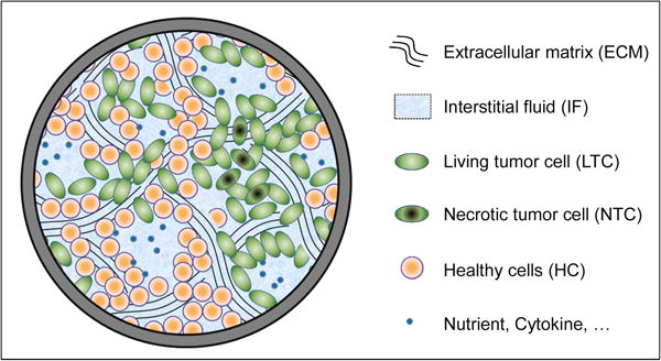
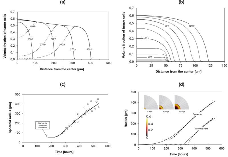
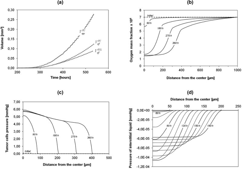
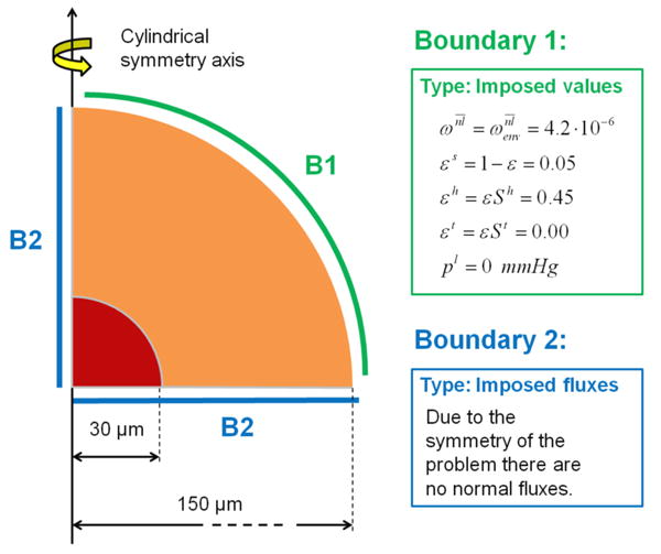
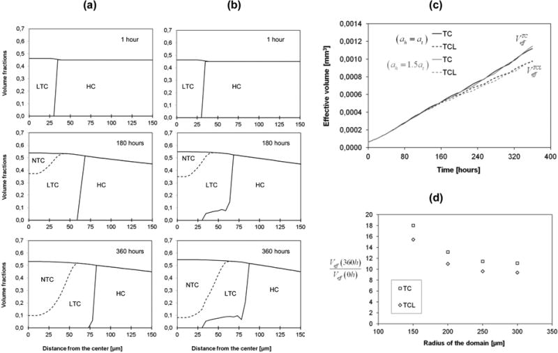
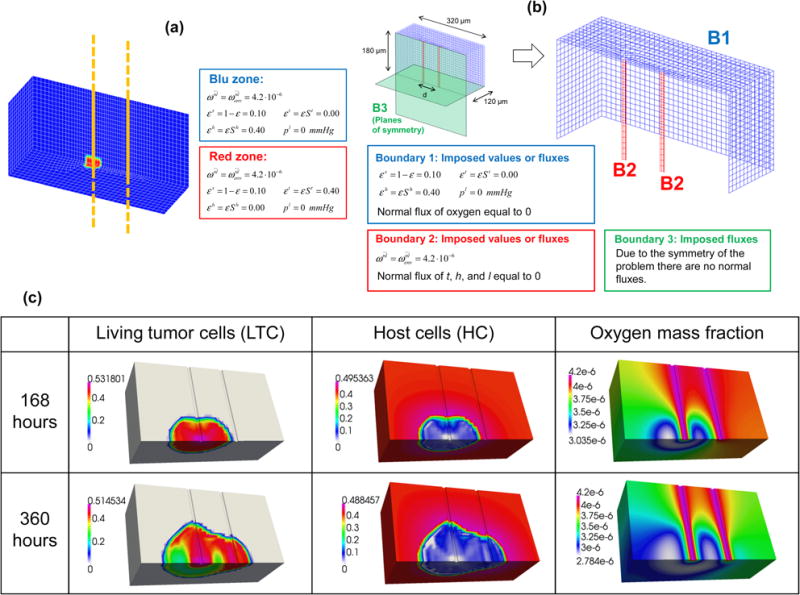
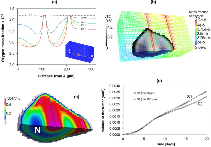
Several mathematical formulations have analyzed the time-dependent behaviour of a tumor mass. However, most of these propose simplifications that compromise the physical soundness of the model. Here, multiphase porous media mechanics is extended to model tumor evolution, using governing equations obtained via the Thermodynamically Constrained Averaging Theory (TCAT). A tumor mass is treated as a multiphase medium composed of an extracellular matrix (ECM); tumor cells (TC), which may become necrotic depending on the nutrient concentration and tumor phase pressure; healthy cells (HC); and an interstitial fluid (IF) for the transport of nutrients. The equations are solved by a Finite Element method to predict the growth rate of the tumor mass as a function of the initial tumor-to-healthy cell density ratio, nutrient concentration, mechanical strain, cell adhesion and geometry. Results are shown for three cases of practical biological interest such as multicellular tumor spheroids (MTS) and tumor cords. First, the model is validated by experimental data for time-dependent growth of an MTS in a culture medium. The tumor growth pattern follows a biphasic behaviour: initially, the rapidly growing tumor cells tend to saturate the volume available without any significant increase in overall tumor size; then, a classical Gompertzian pattern is observed for the MTS radius variation with time. A core with necrotic cells appears for tumor sizes larger than 150 μm, surrounded by a shell of viable tumor cells whose thickness stays almost constant with time. A formula to estimate the size of the necrotic core is proposed. In the second case, the MTS is confined within a healthy tissue. The growth rate is reduced, as compared to the first case - mostly due to the relative adhesion of the tumor and healthy cells to the ECM, and the less favourable transport of nutrients. In particular, for tumor cells adhering less avidly to the ECM, the healthy tissue is progressively displaced as the malignant mass grows, whereas tumor cell infiltration is predicted for the opposite condition. Interestingly, the infiltration potential of the tumor mass is mostly driven by the relative cell adhesion to the ECM. In the third case, a tumor cord model is analyzed where the malignant cells grow around microvessels in a 3D geometry. It is shown that tumor cells tend to migrate among adjacent vessels seeking new oxygen and nutrient. This model can predict and optimize the efficacy of anticancer therapeutic strategies. It can be further developed to answer questions on tumor biophysics, related to the effects of ECM stiffness and cell adhesion on tumor cell proliferation.
Keywords: Finite Elements; cell adhesion; multiphase systems; porous mechanics; tumor cord; tumor growth; tumor spheroid.
Figures
Similar articles
-
Tumor growth modeling from the perspective of multiphase porous media mechanics.Mol Cell Biomech. 2012 Sep;9(3):193-212. Mol Cell Biomech. 2012. PMID: 23285734 Free PMC article.
-
A two-phase model of plantar tissue: a step toward prediction of diabetic foot ulceration.Int J Numer Method Biomed Eng. 2014 Nov;30(11):1153-69. doi: 10.1002/cnm.2650. Epub 2014 May 19. Int J Numer Method Biomed Eng. 2014. PMID: 24841993
-
A tumor growth model with deformable ECM.Phys Biol. 2014 Nov 26;11(6):065004. doi: 10.1088/1478-3975/11/6/065004. Phys Biol. 2014. PMID: 25427284 Free PMC article.
-
Modelling non-homogeneous stochastic reaction-diffusion systems: the case study of gemcitabine-treated non-small cell lung cancer growth.BMC Bioinformatics. 2012;13 Suppl 14(Suppl 14):S14. doi: 10.1186/1471-2105-13-S14-S14. Epub 2012 Sep 7. BMC Bioinformatics. 2012. PMID: 23095709 Free PMC article.
-
Predicting the growth of glioblastoma multiforme spheroids using a multiphase porous media model.Biomech Model Mechanobiol. 2016 Oct;15(5):1215-28. doi: 10.1007/s10237-015-0755-0. Epub 2016 Jan 8. Biomech Model Mechanobiol. 2016. PMID: 26746883
Cited by
-
Osteolytic vs. Osteoblastic Metastatic Lesion: Computational Modeling of the Mechanical Behavior in the Human Vertebra after Screws Fixation Procedure.J Clin Med. 2022 May 18;11(10):2850. doi: 10.3390/jcm11102850. J Clin Med. 2022. PMID: 35628977 Free PMC article.
-
Rapid Biofabrication of an Advanced Microphysiological System Mimicking Phenotypical Heterogeneity and Drug Resistance in Glioblastoma.Adv Healthc Mater. 2024 Dec;13(30):e2401876. doi: 10.1002/adhm.202401876. Epub 2024 Aug 5. Adv Healthc Mater. 2024. PMID: 39101329 Free PMC article.
-
An Eulerian formulation of inelasticity: from metal plasticity to growth of biological tissues.Philos Trans A Math Phys Eng Sci. 2019 May 6;377(2144):20180071. doi: 10.1098/rsta.2018.0071. Philos Trans A Math Phys Eng Sci. 2019. PMID: 30879413 Free PMC article. Review.
-
FDG-PET-based differential uptake volume histograms: a possible approach towards definition of biological target volumes.Br J Radiol. 2016 Jun;89(1062):20150388. doi: 10.1259/bjr.20150388. Epub 2016 Mar 23. Br J Radiol. 2016. PMID: 27007269 Free PMC article.
-
Computational Modelling of Cancer Nanomedicine: Integrating Hyperthermia Treatment Into a Multiphase Porous-Media Tumour Model.Int J Numer Method Biomed Eng. 2025 Aug;41(8):e70074. doi: 10.1002/cnm.70074. Int J Numer Method Biomed Eng. 2025. PMID: 40765034 Free PMC article.
References
-
- Ambrosi D, Preziosi L, Vitale G. The interplay between stress and growth in solid tumors. Mech Res Comm. 2012;42:87–91.
-
- Anderson AR. A hybrid mathematical model of solid tumour invasion: the importance of cell adhesion. Math Med Biol. 2005;22:163–186. - PubMed
-
- Astanin S, Preziosi L. Mathematical modelling of the Warburg effect in tumour cords. J Theor Biol. 2009;258(4):578–590. - PubMed
Grants and funding
LinkOut - more resources
Full Text Sources
Other Literature Sources
Miscellaneous