

Porcine incisional hernia model: Evaluation of biologically derived intact extracellular matrix repairs
- PMID: 24555008
- PMCID: PMC3927864
- DOI: 10.1177/2041731413508771
Porcine incisional hernia model: Evaluation of biologically derived intact extracellular matrix repairs
Abstract
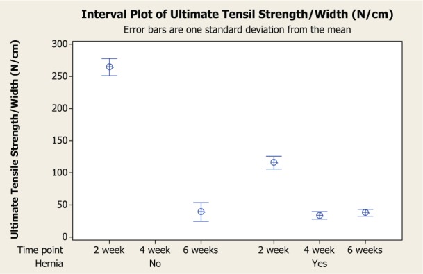
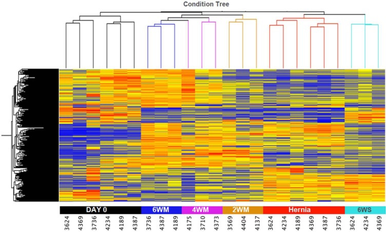
We compared fascial wounds repaired with non-cross-linked intact porcine-derived acellular dermal matrix versus primary closure in a large-animal hernia model. Incisional hernias were created in Yucatan pigs and repaired after 3 weeks via open technique with suture-only primary closure or intraperitoneally placed porcine-derived acellular dermal matrix. Progressive changes in mechanical and biological properties of porcine-derived acellular dermal matrix and repair sites were assessed. Porcine-derived acellular dermal matrix-repaired hernias of additional animals were evaluated 2 and 4 weeks post incision to assess porcine-derived acellular dermal matrix regenerative potential and biomechanical changes. Hernias repaired with primary closure showed substantially more scarring and bone hyperplasia along the incision line. Mechanical remodeling of porcine-derived acellular dermal matrix was noted over time. Porcine-derived acellular dermal matrix elastic modulus and ultimate tensile stress were similar to fascia at 6 weeks. The biology of porcine-derived acellular dermal matrix-reinforced animals was more similar to native abdominal wall versus that with primary closure. In this study, porcine-derived acellular dermal matrix-reinforced repairs provided more complete wound healing response compared with primary closure.
Keywords: Incisional hernia; Strattice; biologic mesh; biologic tissue matrix; fascial wounds.
Conflict of interest statement
Figures


Similar articles
-
Non-cross-linked porcine acellular dermal matrices for abdominal wall reconstruction.Plast Reconstr Surg. 2010 Jan;125(1):167-176. doi: 10.1097/PRS.0b013e3181c2a6ed. Plast Reconstr Surg. 2010. PMID: 19910855
-
Primary fascial closure with biologic mesh reinforcement results in lesser complication and recurrence rates than bridged biologic mesh repair for abdominal wall reconstruction: A propensity score analysis.Surgery. 2017 Feb;161(2):499-508. doi: 10.1016/j.surg.2016.08.009. Epub 2016 Oct 31. Surgery. 2017. PMID: 27810091
-
Evaluation of human acellular dermis versus porcine acellular dermis in an in vivo model for incisional hernia repair.Cell Tissue Bank. 2011 May;12(2):135-45. doi: 10.1007/s10561-011-9245-5. Epub 2011 Mar 6. Cell Tissue Bank. 2011. PMID: 21380733 Free PMC article.
-
Human acellular dermal matrix for repair of abdominal wall defects: review of clinical experience and experimental data.J Long Term Eff Med Implants. 2005;15(5):547-58. doi: 10.1615/jlongtermeffmedimplants.v15.i5.70. J Long Term Eff Med Implants. 2005. PMID: 16218902 Review.
-
Porcine acellular dermal matrix (PADM) vascularises after exposure in open necrotic wounds seen after complex hernia repair.Int Wound J. 2016 Oct;13(5):972-6. doi: 10.1111/iwj.12558. Epub 2015 Dec 21. Int Wound J. 2016. PMID: 26688300 Free PMC article. Review.
Cited by
-
Comparison of mechanical properties and host tissue response to OviTex™ and Strattice™ surgical meshes: author reply.Hernia. 2024 Feb;28(1):281-282. doi: 10.1007/s10029-023-02911-y. Epub 2023 Oct 19. Hernia. 2024. PMID: 37855939 Free PMC article. No abstract available.
-
Comparison of mechanical properties and host tissue response to OviTex™ and Strattice™ surgical meshes.Hernia. 2023 Aug;27(4):987-997. doi: 10.1007/s10029-023-02769-0. Epub 2023 Apr 8. Hernia. 2023. PMID: 37031315 Free PMC article.
-
Evaluation of a fully absorbable poly-4-hydroxybutyrate/absorbable barrier composite mesh in a porcine model of ventral hernia repair.Surg Endosc. 2016 Sep;30(9):3691-701. doi: 10.1007/s00464-016-5057-9. Epub 2016 Jul 1. Surg Endosc. 2016. PMID: 27369286 Free PMC article.
-
Collagen Remodeling of Strattice™ Firm in a Nonhuman Primate Model of Abdominal Wall Repair.Bioengineering (Basel). 2025 Jul 24;12(8):796. doi: 10.3390/bioengineering12080796. Bioengineering (Basel). 2025. PMID: 40868309 Free PMC article.
References
-
- Luijendijk RW, Hop WC, van den Tol MP, et al. A comparison of suture repair with mesh repair for incisional hernia. N Engl J Med 2000; 343(6): 392–398 - PubMed
-
- Vrijland WW, van den Tol MP, Luijendijk RW, et al. Randomized clinical trial of non-mesh versus mesh repair of primary inguinal hernia. Br J Surg 2002; 89(3): 293–297 - PubMed
-
- Boruch AV, Nieponice A, Qureshi IR, et al. Constructive remodeling of biologic scaffolds is dependent on early exposure to physiologic bladder filling in a canine partial cystectomy model. J Surg Res 2010; 161(2): 217–225 - PubMed
-
- Bellows CF, Alder A, Helton WS. Abdominal wall reconstruction using biological tissue grafts: present status and future opportunities. Expert Rev Med Devices 2006; 3(5): 657–675 - PubMed
LinkOut - more resources
Full Text Sources
Other Literature Sources
