

doi: 10.1155/2012/374107.
Epub 2012 Jul 12.
Retroperitoneal extrarenal angiomyolipomas: an evidence-based approach to a rare clinical entity
Affiliations
- PMID: 24555133
- PMCID: PMC3914176
- DOI: 10.1155/2012/374107
Item in Clipboard
Retroperitoneal extrarenal angiomyolipomas: an evidence-based approach to a rare clinical entity
Case Rep Nephrol.
2012.
Abstract
Extrarenal angiomyolipomas (ERAMLs) are rare tumors that present as incidentalomas upon imaging for other conditions. Retroperitoneal ERAMLs present a unique challenge from a diagnostic and treatment standpoint as they can mimic other benign and malignant retroperitoneal tumors. We present a case of a 39-year-old female with a 19.3 cm × 13.5 cm × 10.7 cm left extrarenal retroperitoneal mass. Histopathologic examination and HMB-45 staining revealed the mass to be a retroperitoneal ERAML. Our case report provides a comprehensive literature review and an evidence-based algorithm for taking care of patients with ERAMLs.
Figures

(a) oral contrast and (b) IV and oral contrast: abdominal computerized tomography demonstrating an encapsulated fatty vascular mass (white arrows) lateral to the left kidney measuring 19.3 cm × 13.5 cm × 10.7 cm with prominent vascular dependence on the left renal vein and artery as well as a 2 cm posterior mid-pole homogeneous fatty density (yellow arrow). Left colon is laterally displaced (orange arrow).

Abdominal magnetic resonance imaging demonstrating a large fatty encapsulated mass (white asterisk) measuring 19.3 cm × 13.5 cm × 10.7 cm with prominent vascularity (white arrows). The anatomic relationship between the mass and the left kidney can be well seen in Figure 2(b).

Gross image of the en bloc resected mass including the left kidney (black arrow), demonstrating a well-encapsulated fatty mass attached to the upper pole of the kidney (white arrow) with a smooth outer surface measuring 23 cm × 14 cm × 9 cm.
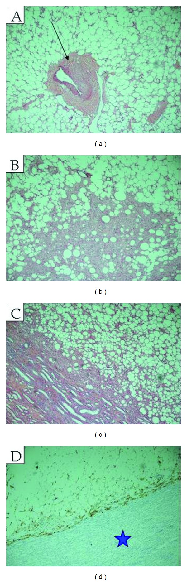
Extrarenal mass (Hematoxylin and Eosin). Photomicrograph of the mass demonstrate mature adipose tissue with a tortuous thick blood vessel (black arrow) ((a); ×20) and bundles of smooth muscles lacking elastic tissue lamina ((b); ×40), adipose tissue with small areas of smooth muscle with epithelioid features ((c); ×40). Focal staining with HMB45 antibody was positive (blue star) ((d); ×40), consistent with angiomyolipoma.
References
-
- Friis J, Hjortrup A. Extrarenal angiomyolipoma: diagnosis and management. Journal of Urology. 1982;127(3):528–529. - PubMed
-
- Tsutsumi M, Yamauchi A, Tsukamoto S, Ishikawa S. A case of angiomyolipoma presenting as a huge retroperitoneal mass. International Journal of Urology. 2001;8(8):470–471. - PubMed
-
- Demopoulos RI, Denarvaez F, Kaji V. Benign mixed mesodermal tumors of the uterus: a histogenetic study. American Journal of Clinical Pathology. 1973;60(3):377–383. - PubMed
-
- Gutmann J, Cifuentes C, Vicuna R, Sobarzo V, Balzarini MA. Intraoral angiomyolipoma. Oral Surgery, Oral Medicine, Oral Pathology. 1975;39(6):945–948. - PubMed
-
- Bures C, Barnes L. Benign mesenchymomas of the head and neck. Archives of Pathology and Laboratory Medicine. 1978;102(5):237–241. - PubMed
LinkOut - more resources
Full Text Sources
