

Case Reports
doi: 10.15274/INR-2014-10009.
Epub 2014 Feb 10.
The donut sign: a new angiographic sign for partially thrombosed aneurysms with flow-induced intraluminal thrombus
Affiliations
- PMID: 24556300
- PMCID: PMC3971142
- DOI: 10.15274/INR-2014-10009
Item in Clipboard
Case Reports
The donut sign: a new angiographic sign for partially thrombosed aneurysms with flow-induced intraluminal thrombus
Interv Neuroradiol.
2014 Jan-Feb.
Abstract
Three patients are described with unruptured large partially thrombosed aneurysms with a peculiar donut-shaped remaining lumen. Observations suggest that the flow geometry of the aneurysm and parent vessels induces a preferential circular laminar flow inside the aneurysm followed by partial intraluminal thrombosis leaving a donut-shaped lumen to accommodate the circular flow. This flow mechanism of thrombus formation inside aneurysms is different from the more common repeated intramural dissections and hemorrhages that cause growth in most large and giant partially thrombosed aneurysms.
Keywords: 3D rotational angiography; intracranial aneurysm; partial thrombosis.
Figures
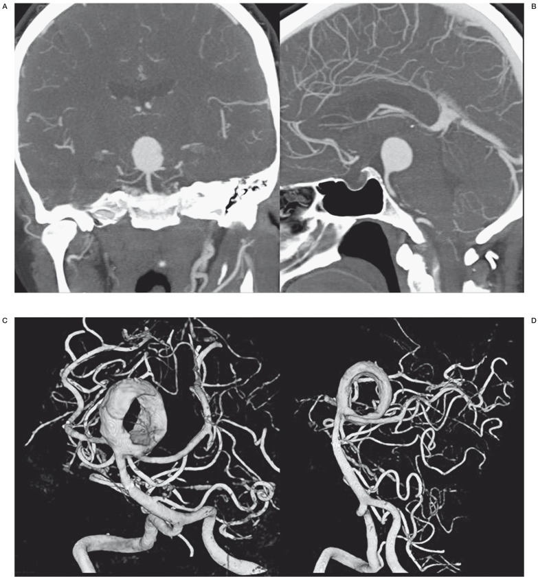
Incidental large basilar tip aneurysm in a 50-year-old woman. A,B) CT angiography demonstrates large spherical basilar tip aneurysm. C,D) 3DRA of the same aneurysm 10 days after stent placement in the right posterior cerebral artery. The aneurysm is now largely thrombosed with a donut-shaped remaining lumen.
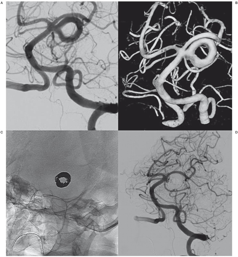
Incidental superior cerebellar artery aneurysm in a 70-year-old woman. A,B) 2D angiography (A) and 3DRA (B) show the large left superior cerebellar artery aneurysm with donut–shaped lumen. C) Circular deposition of coils in the donut-shaped lumen. D) Almost complete occlusion after coiling.
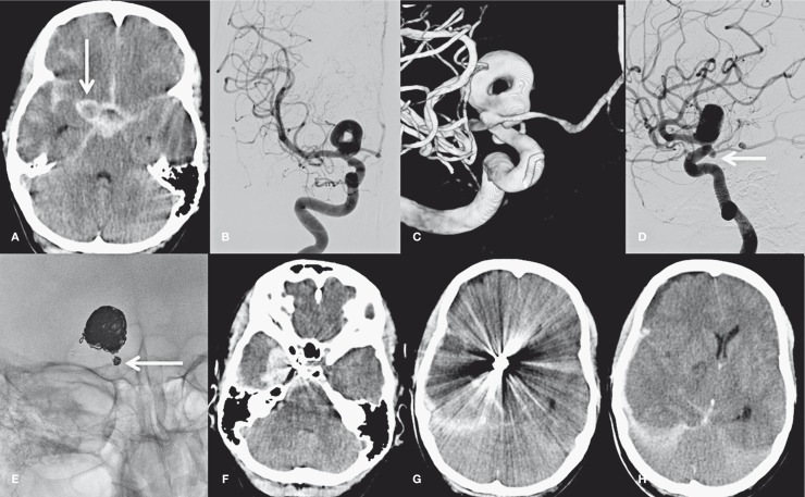
A 46-year-old man with poor grade subarachnoid hemorrhage. A) CT scan shows SAH and aneurysm (arrow). B-D) 2D angiography (B,D) and 3DRA (C) demonstrate a donut-shaped large carotid tip aneurysm and a second aneurysm on the PcomA (arrow in D). E) Coil meshes after coiling of the carotid tip aneurysm and the small PcomA aneurysm (arrow). F-H) CT scan 10 days after coiling shows recurrent hemorrhage from the PcomA aneurysm with a large subdural component and mass effect.
References
-
- Martin AJ, Hetts SW, Dillon WP, et al. MR imaging of partially thrombosed cerebral aneurysms: characteristics and evolution. Am J Neuroradiol. 2011;32:346–351. doi: 10.3174/ajnr.A2298. - DOI - PMC - PubMed
-
- Roccatagliata L, Guédin P, Condette-Auliac S, et al. Partially thrombosed intracranial aneurysms: symptoms, evolution, and therapeutic management. Acta Neurochir (Wien) 2010;152:2133–2142. doi: 10.1007/s00701-010-0772-9. - DOI - PubMed
-
- Ferns SP, van Rooij WJ, Sluzewski M, et al. Partially thrombosed intracranial aneurysms presenting with mass effect: long-term clinical and imaging follow-up after endovascular treatment. Am J Neuroradiol. 2010;31:1197–1205. doi: 10.3174/ajnr.A2057. - DOI - PMC - PubMed
-
- van Rooij WJ, Sluzewski M, Beute GN. Endovascular treatment of giant serpentine aneurysms. Am J Neuroradiol. 2008;29:1418–1419. doi: 10.3174/ajnr.A1071. - DOI - PMC - PubMed
Publication types
MeSH terms
LinkOut - more resources
Full Text Sources
Other Literature Sources
Medical
