Review
. 2014 Sep:182:3-18.
doi: 10.1016/j.chemphyslip.2014.02.002. Epub 2014 Feb 17.

Cellular and molecular interactions of phosphoinositides and peripheral proteins

Affiliations
Review

Cellular and molecular interactions of phosphoinositides and peripheral proteins

Robert V Stahelin et al. Chem Phys Lipids. 2014 Sep.

Abstract

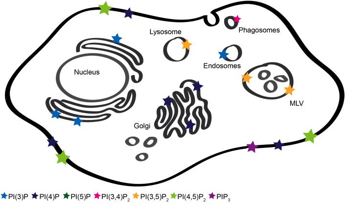
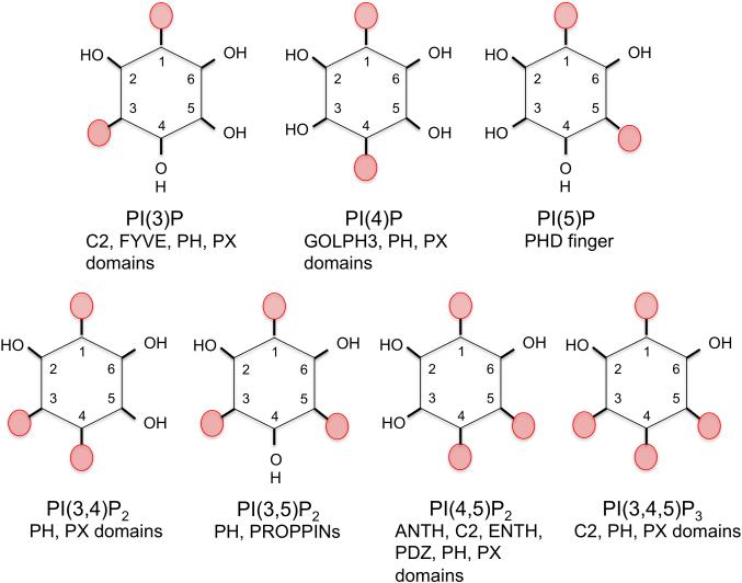
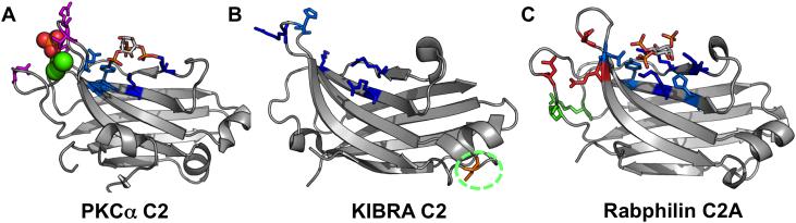
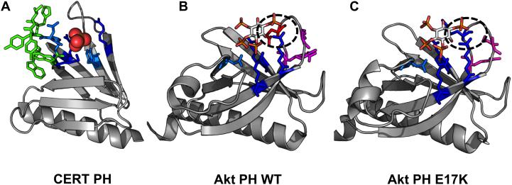
Anionic lipids act as signals for the recruitment of proteins containing cationic clusters to biological membranes. A family of anionic lipids known as the phosphoinositides (PIPs) are low in abundance, yet play a critical role in recruitment of peripheral proteins to the membrane interface. PIPs are mono-, bis-, or trisphosphorylated derivatives of phosphatidylinositol (PI) yielding seven species with different structure and anionic charge. The differential spatial distribution and temporal appearance of PIPs is key to their role in communicating information to target proteins. Selective recognition of PIPs came into play with the discovery that the substrate of protein kinase C termed pleckstrin possessed the first PIP binding region termed the pleckstrin homology (PH) domain. Since the discovery of the PH domain, more than ten PIP binding domains have been identified including PH, ENTH, FYVE, PX, and C2 domains. Representative examples of each of these domains have been thoroughly characterized to understand how they coordinate PIP headgroups in membranes, translocate to specific membrane docking sites in the cell, and function to regulate the activity of their full-length proteins. In addition, a number of novel mechanisms of PIP-mediated membrane association have emerged, such as coincidence detection-specificity for two distinct lipid headgroups. Other PIP-binding domains may also harbor selectivity for a membrane physical property such as charge or membrane curvature. This review summarizes the current understanding of the cellular distribution of PIPs and their molecular interaction with peripheral proteins.

Keywords: C2 domain; FYVE domain; Lipid binding; Membrane binding; PH domain; PI(3)P; PI(3,4,5)P(3); PI(4,5)P(2); Peripheral protein.; Phosphoinsoitide.


Figures

Figure 1
Figure 1. Cellular Distribution of PIPs
A cartoon of a cell is shown to illustrate the known location of PIPs in mammalian cells. The relative abundance of PIPs is depicted with the size of the star shown.
Figure 2
Figure 2. There are seven different PIPs in mammalian cells that mediate recruitment of peripheral proteins to cellular membranes
The headgroup of each PI is shown with known effector domains listed below each.
Figure 3
Figure 3. C2 domains have a β-sandwich structure with loop regions adjacent to the “cationic patch”
These structures have been crystallized with or without inositol headgroups and demonstrate PI-binding through interaction with positively-charged residues in the cationic patch region. PI-binding residues are depicted in blue. Dark blue residues represent positively charged lysines or argininges and light blue are other inositol-headgroup coordinating residues. Residues highlighted in magenta are PS-binding, residues highlighted in green are membrane-inserting, and residues highlighted in red are negative and calcium-coordinating. A. PKCα C2 (3GPE) coordinates PS with Asn189, Arg216, Arg249, and Thr251 where the sulfate is coordinated in the structure (orange and red spheres). In this loop region, negatively charged residues coordinate 3 calcium inos (green spheres) which are crucial to its membrane localization. The PIP2 headgroup is coordinated by Tyr195, Lys197, Lys209, Lys221, Trp245, and Asn253 in the cationic patch region. B. KIBRA C2 (2ZOU) harbors a cationic patch region with Arg753, His755, Arg714, Arg716, Lys692, and Arg696. In addition, Cys771 participates in a disulfide bond between two monomers in the structure. While the molecular basis of WT KIBRA binding to monophosphorylated PIPs such as PI(3)P is unknown, mutation of Met734 or Ser735 alters the C2 domain affinity in such a way it increases the affinity of the C2 domains for PI(4)P, PI(5)P, and PI(4,5)P2 (Duning et al. 2013). C. Rabphilin C2A bound to IP3 (4NP9) coordinates the inositol headgroup in the cationic patch region using Tyr421, Lys423, His425, Lys435, Arg437 and Asn481.
Figure 4
Figure 4. Pleckstrin Homology (PH) domains have a β-barrel structure with an alpha helical cap
These structures have been crystallized with either sulfate of inositol headgroups and demonstrate PI-binding at one end of the barrel with positively-charged residues and hydrophobic residues which insert into the membrane from the loop regions of the protein. PI-binding residues are depicted in blue. Dark blue residues are positively charged lysines or argininges and light blue are other inositol-headgroup coordinating residues. Residues highlighted in green insert themselves into the membrane from the loop regions, and residues highlighted in magenta are PS-binding. A. CERT PH (4HHV) coordinates a sulfate ion (orange and yellow spheres) in the inositol-binding region with Lys32, Thr34, Asn35, Arg43, Tyr54, Arg66 and inserts into the membrane with Trp33, Tyr36, Ile37, His38, Trp40. B. Akt PH (1UNQ) coordinates IP4 with Glu17, Arg23, Arg25, Asn52, and Arg86. It coordinates PS with Arg15 and Lys20. C. Akt PH (2UZS) is known to be converted to a constitutively active form in some cancers via mutation of Glu17 to Lys17 (highlighted by the black ovals in B and C).

References

    1. Adu-Gyamfi E, Soni SP, Xue Y, Digman MA, Gratton E, Stahelin RV. The Ebola virus matrix protein penetrates into the plasma membrane: a key step in viral protein 40 (VP40) oligomerization and viral egress. J. Biol. Chem. 2013;288:5779–5789. - PMC - PubMed
    1. Agranoff BW, Bradley RM, Brady RO. The enzymatic synthesis of inositol phosphatide. J. Biol. Chem. 1958;233:1077–1083. - PubMed
    1. Alajlouni R, Drahos KE, Finkielstein CV, Capelluto DG. Lipid-mediated membrane binding properties of Disabled-2. Biochim. Biophys. Acta. 2011;1808:2734–2744. - PubMed
    1. Ankem G, Mitra S, Sun F, Moreno AC, Chutvirasakul B, Azurmend HF, Li L, Capelluto DGS. The C2 domain of Tollip, a toll-like receptor signaling regulator, exhibits broad preference for phosphoinositides. Biochem. J. 2011;435:597–608. - PubMed
    1. Arai Y, Ijuin T, Takenawa T, Becker LE, Takashima S. Excessive expression of synaptojanin in brains with Down syndrome. Brain Development. 2002;24:67–72. - PubMed

Publication types