

The central role of antigen presentation in islets of Langerhans in autoimmune diabetes
- PMID: 24556398
- PMCID: PMC4118295
- DOI: 10.1016/j.coi.2013.10.011
The central role of antigen presentation in islets of Langerhans in autoimmune diabetes
Abstract
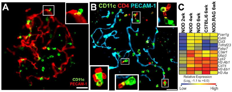
The islets of Langerhans normally contain resident antigen presenting cells (APCs), which in normal conditions are mostly represented by macrophages, with a few dendritic cells (DC). We present here the features of these islet APCs, making the point that they have a supportive function in islet homeostasis. Islet APCs express high levels of major histocompatibility complexes (MHC) molecules on their surfaces and are highly active in antigen presentation in the autoimmune diabetes of the NOD mouse: they do this by presenting peptides derived from molecules of the β-cells. These APCs also are instrumental in the localization of diabetogenic T cells into islets. The islet APC present exogenous peptides derived from secretory granules of the β-cell, giving rise to unique peptide-MHC complexes (pMHC) that activate those non-conventional T cells that bypass thymus selection.
Copyright © 2013 Elsevier Ltd. All rights reserved.
Figures
References
-
- Lacy PE, Davie JM, Finke EH. Prolongation of islet allograft survival following in vitro culture (24 degrees C) and a single injection of ALS. Science. 1979;204:312–316. - PubMed
-
- Simeonovic CJ, Bowen KM, Kotlarski I, Lafferty KJ. Modulation of tissue immunogenicity by organ culture. Comparison of adult islets and fetal pancreas. Transplantation. 1980;30:174–179. - PubMed
-
- Snell GD. The homograft reaction. Ann Rev Microbiol. 1957;11:439–458. - PubMed
-
- Lafferty KJ, Prowse SJ, Simeonovic CJ. Immunobiology of tissue transplantation: A return to the passenger leukocyte concept. Ann Rev Immunol. 1983;1:143–173. Ref. 2,3, and 5 are early papers reporting APC in islets of Langerhans. - PubMed
Publication types
MeSH terms
Substances
Grants and funding
LinkOut - more resources
Full Text Sources
Other Literature Sources
Medical
Research Materials
Miscellaneous
