Review
. 2014 Feb:26:100-10.
doi: 10.1016/j.coi.2013.11.003. Epub 2013 Dec 5.

Insights into phagocytosis-coupled activation of pattern recognition receptors and inflammasomes

Affiliations
Review

Insights into phagocytosis-coupled activation of pattern recognition receptors and inflammasomes

Julien Moretti et al. Curr Opin Immunol. 2014 Feb.

Abstract

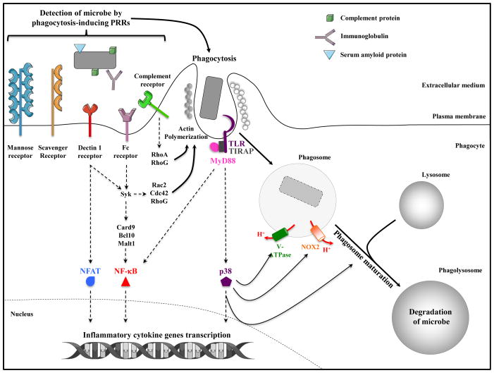
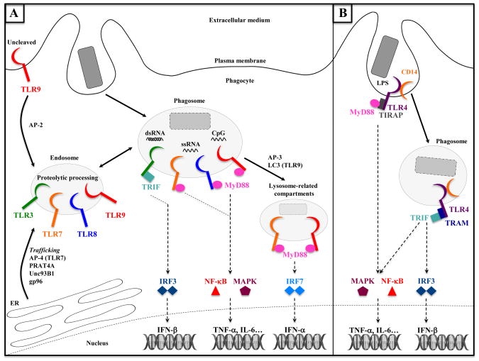
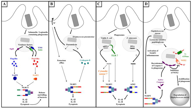
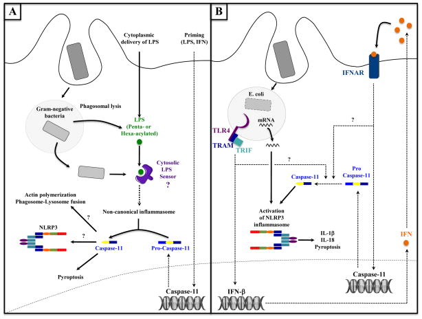
A decade of work shows that the core function of phagocytosis in engulfment and destruction of microorganisms is only a small facet of the full spectrum of roles for phagocytosis in the immune system. The regulation of phagocytosis and its outcomes by inflammatory pattern recognition receptors (PRRs) is now followed by new studies strengthening this concept and adding further complexity to the relationship between phagocytosis and innate immune signaling. Phagocytosis forms the platform for activation of distinct members of the Toll-like receptor family, and even dictates their signaling outcomes. In many cases, phagocytosis is a necessary precedent to the activation of cytosolic PRRs and assembly of canonical and non-canonical inflammasomes, leading to strong pro-inflammatory responses and inflammatory cell death.


Figures

Figure 1
Figure 1. Signal-dependent induction of phagocytosis and phagosome maturation
Phagocytosis is an actin-driven, receptor mediated process initiated upon recognition of microorganisms by Pattern Recognition Receptors (PRRs) expressed at the plasma membrane of phagocytes. Microorganisms can also be opsonized by immunoglobulins, serum amyloid P component or complement proteins, which engage specific PRRs and trigger opsonic phagocytosis. Some receptors like scavenger receptors and mannose receptor serve as phagocytic PRRs, while others like Dectin-1 and FcγR serve dual roles transmitting inflammatory signals receptor that activate NF-κB and/or NFAT transcription factors, and triggering actin polymerization via Rac2, Cdc42, and RhoG [75]. Toll-like receptors (TLRs) are signaling PRRs engaged by microbial components during phagocytosis leading to the activation of NF-κB, MAPK and other transcription factors. TLR signals also trigger an inducible rate of phagocytosis and phagosome maturation via MyD88-dependent activation of the MAPK p38. Signals from TLRs also induce assembly of the NADPH oxidase NOX2 and the vacuolar v-ATPase.
Figure 2
Figure 2. The phagosome is a signaling compartment for TLRs: cases of endosomal TLRs and TLR4
(A) TLR3, TLR7, TLR8 and TLR9 are endosomal receptors. Trafficking of these receptors from the endoplasmic reticulum (ER) to endosomes requires chaperone proteins such as UNC93B1, gp96, AP-4 (for trafficking TLR7 directly to endosomes) or AP-2 (for trafficking TLR9 from the intermediate plasma membrane step between ER and endosomes). Microbial nucleic acids released upon degradation of microorgamisms activate endosomal TLRs delivered to phagosomes. TLR3 is activated by dsRNA and signals through TRIF to activate IRF3 and IFN-β transcription. TLR7, TLR8 (both activated by ssRNA) and TLR9 (sensing CpG DNA) signal through MyD88 and activate NF-κB and MAPK signaling pathways, leading to the transcription of genes such as TNF-α or IL-6. AP-3 and LC3 enable trafficking and signaling of TLR7 and TLR9 to lysosome-related compartments, where these receptors specifically activate the transcription of IFN-α genes in a MyD88- and IRF7-dependent manner. (B) At the plasma membrane, TLR4 recognizes LPS and signals through MyD88 to activate NF-κB and MAPK. CD14 mediates the endocytosis of TLR4, which undergoes a signaling switch from plasma membrane MyD88 to endosomal/phagosomal TRIF signaling and activation of the IRF3-dependent transcription of IFN-β genes.
Figure 3
Figure 3. Phagocytosis enables activation of inflammasomes
A. Activation of the NLRC4 inflammasome through the activity of microbial virulence factors. Phagocytosed Salmonella or Legionella inject proteins into the cytosol of phagocytes via type III and type IV secretion systems (T3SS and T4SS, respectively) or associated translocon proteins such as SipB (from T3SS). Flagellin or PrgJ-like proteins (T3SS rod proteins) are released into the cytosol where they activate the NLRC4 inflammasome. Flagellin binding to NAIP5 and PrgJ binding to NAIP2 are required for NLRC4 activation. Inflammasome-dependent pyroptosis leads to the release of bacteria from macrophages and their subsequent phagocytosis and killing by neutrophils (also the case in panels B, C and D). B. Activation of the NLRP3 inflammasome by the pore-forming toxin Pneumolysin. Phagocytosed Streptococcus pneumoniae secrete the pore-forming toxin Pneumolysin in phagosomes inducing disruption of phagosomal membranes. Subsequent potassium efflux, or release of the lysosomal protease Cathepsin B into the cytosol can lead to activation of the NLRP3 inflammasome. C. Activation of NLRP3 and AIM2 inflammasomes by microbial nucleic acids. E. coli mRNA is a signature of microbial viability, which is detected upon or after phagocytosis of bacteria (potentially via a TLR or cytosolic RNA sensor), activates the NLRP3 inflammasome, and triggers IL-1β and IL-18 production and pyroptosis. Phagocytosis of F. tularensis phagocytosis leads to access of bacterial DNA to the cytosol where it activates the AIM2 inflammasome. D. Interaction between phagosome and NLRP3 inflammasome. The NLRP3 inflammasome can be activated upon phagocytosis of Staphylococcus aureus when its PGN lacks modifications rendering it sensitive to lysozyme degradation. Active caspase-1 has been demonstrated around Staphylococcus-containing phagosomes, where it inhibits the NADPH oxidase NOX2. As a consequence, phagosomes acidify and Staphylococcus degradation in phagosomes is enhanced.
Figure 4
Figure 4. Phagocytosis and activation of non-canonical inflammasome and caspase-11
A. Activation of the non-canonical inflammasome after phagosomal lysis and leaking of bacterial components. Direct delivery of LPS into the cytosol either experimentally with cholera toxin B or via transfection, or physiologically upon infection with Gram-negative bacteria, enables its detection by an unidentified cytosolic LPS-sensor. This sensor detects specifically hexa- or penta-acylated LPS, and then activates the non-canonical inflammasome pathway and caspase-11 within a few hours following infection. Active caspase-11 then promotes pyroptosis, and could also participate in the activation of the NLRP3 inflammasome or regulate phagosome-lysosome fusion [76]. Note that a priming step is required to induce caspase-11 transcription. B. Activation of caspase-11 and its role in activation of the NLRP3 inflammasome. Upon phagocytosis of viable E. coli, microbial mRNA induces the assembly and activation of the NLRP3 inflammasome, while TRIF-dependent TLR4 signaling activates the transcription of IFN-β genes. Secreted IFN-β binds to the type I IFN receptor (IFNAR), which induces the transcription of caspase-11 gene. Pro-caspase-11 is then cleaved into active caspase-11 and synergizes with bacterial mRNA in NLRP3 inflammasome. Note that TRIF and IFNAR signaling are involved in late stage (12–16 hours post infection) caspase-11 activation via as yet unidentified steps.

References

    1. Torchinsky MB, Garaude J, Blander JM. Infection and apoptosis as a combined inflammatory trigger. Curr Opin Immunol. 2010;22:55–62. - PMC - PubMed
    1. Nair P, Amsen D, Blander JM. Co-ordination of incoming and outgoing traffic in antigen-presenting cells by pattern recognition receptors and T cells. Traffic. 2011;12 :1669–1676. - PMC - PubMed
    1. Takeuchi O, Akira S. Pattern recognition receptors and inflammation. Cell. 2010;140:805–820. - PubMed
    1. Blander JM. Signalling and phagocytosis in the orchestration of host defence. Cell Microbiol. 2007;9:290–299. - PubMed
    1. Underhill DM, Goodridge HS. Information processing during phagocytosis. Nat Rev Immunol. 2012;12:492–502. - PMC - PubMed

Publication types

MeSH terms

LinkOut - more resources