. 2014 Apr 28;180(100):71-80.
doi: 10.1016/j.jconrel.2014.02.007. Epub 2014 Feb 17.

Design and physicochemical characterisation of novel dissolving polymeric microneedle arrays for transdermal delivery of high dose, low molecular weight drugs

Affiliations

Design and physicochemical characterisation of novel dissolving polymeric microneedle arrays for transdermal delivery of high dose, low molecular weight drugs

Maelíosa T C McCrudden et al. J Control Release. .

Abstract

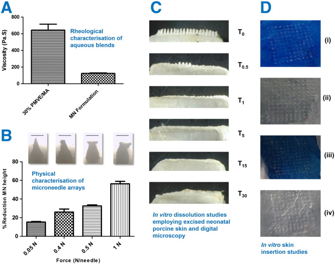
We describe formulation and evaluation of novel dissolving polymeric microneedle (MN) arrays for the facilitated delivery of low molecular weight, high dose drugs. Ibuprofen sodium was used as the model here and was successfully formulated at approximately 50% w/w in the dry state using the copolymer poly(methylvinylether/maleic acid). These MNs were robust and effectively penetrated skin in vitro, dissolving rapidly to deliver the incorporated drug. The delivery of 1.5mg ibuprofen sodium, the theoretical mass of ibuprofen sodium contained within the dry MN alone, was vastly exceeded, indicating extensive delivery of the drug loaded into the baseplates. Indeed in in vitro transdermal delivery studies, approximately 33mg (90%) of the drug initially loaded into the arrays was delivered over 24h. Iontophoresis produced no meaningful increase in delivery. Biocompatibility studies and in vivo rat skin tolerance experiments raised no concerns. The blood plasma ibuprofen sodium concentrations achieved in rats (263μgml(-1) at the 24h time point) were approximately 20 times greater than the human therapeutic plasma level. By simplistic extrapolation of average weights from rats to humans, a MN patch design of no greater than 10cm(2) could cautiously be estimated to deliver therapeutically-relevant concentrations of ibuprofen sodium in humans. This work, therefore, represents a significant progression in exploitation of MN for successful transdermal delivery of a much wider range of drugs.

Keywords: Biocompatibility; Ibuprofen; Microneedles; Transdermal.


Figures

None
Graphical abstract
Fig. 1
Fig. 1
Schematic illustration of the mechanism of drug delivery from dissolving microneedle arrays with containing ibuprofen sodium (A). Digital image of the optimised formulation for dissolving microneedles containing ibuprofen sodium (B). Texture Analyser/light microscopy set-up for investigation of physical properties of microneedles (C) and Franz cell set-up for in vitro transdermal drug release studies (D). Indication of biological testing of microneedles in 3D (E) and 2D (F) cell culture models.
Fig. 2
Fig. 2
Viscosity of 30% w/w PMVE/MA, pH 7.0 gel formulation and the 70% PMVE/MA 30% w/w gel, pH 7.0:30% ibuprofen sodium formulation (optimised microneedle formulation), (means ± S.D., n = 5) (A). Digital microscope images of dissolving PMVE/MA microneedles loaded with ibuprofen sodium following the application of different forces (0.05, 0.4, 0.5 and 1.0 N/needle). Microneedle arrays were attached to the moveable cylindrical probe (length 5.0 cm, cross-sectional area 1.5 cm2) of the Texture Analyser using double-sided adhesive tape and an axial compression load was then applied. The test station pressed the MN arrays against a flat block of aluminium at a rate of 0.5 mm s− 1 with defined forces for 30 s. The pre- and post-test speeds were 1.0 mm s− 1 and the trigger force was set at 0.049 N. These images are representative of the percentage reduction in the heights of needles on the MN arrays observed following the application of the different forces (means + S.D., n = 3). The scale bars represent a length of 300 μm (B). Representative digital micrographs illustrative of the dissolution of ibuprofen sodium-loaded dissolving microneedle arrays (prepared from gels comprised of 70% PMVE/MA 30% w/w gel, pH 7.0:30% ibuprofen sodium) at specific time points (T0: 0 min; T0.5: 0.5 min; T1: 1 min etc.) over a 30 min period following insertion into, and removal from excised neonatal porcine skin (C). Digital images showing microconduits on methylene-blue stained neonatal porcine skin after application of forces of (i) 0.4 & (iii) 0.5 N/needle to ibuprofen sodium-loaded dissolving PMVE/MA microneedle arrays. The images presented in (ii) and (iv) are the reciprocal images of the etching visible on the laboratory film (Parafilm®) lying under the microneedle arrays following force application (D).
Fig. 3
Fig. 3
Percentage recovery of ibuprofen sodium from dissolving PMVE/MA microneedle arrays (means ± S.D., n = 6) (A). The in vitro cumulative permeation profile of ibuprofen sodium across dermatomed 350 μm neonatal porcine skin when delivered using in-dwelling dissolving PMVE/MA microneedle arrays (means ± S.D., n = 6) (B). The inset chart shows the mass balance following completion of the 24 h delivery experiment as compared to the theoretical loading of ibuprofen sodium. The in vitro cumulative permeation profile of ibuprofen-sodium across dermatomed 350 μm neonatal porcine skin when combining iontophoresis (0.5 mA cm− 2) and in-dwelling dissolving PMVE/MA microneedle arrays for a period of 6 h (MN + IP), or passive delivery from dissolving microneedle arrays only (MN) (means ± S.D., n = 5) (C). The in vitro cumulative permeation profile of ibuprofen-sodium across dermatomed 350 μm neonatal porcine skin when combining iontophoresis (0.5 mA cm− 2) and in-dwelling dissolving PMVE/MA microneedle arrays for a period of 30 min (MN + IP), or passive delivery from dissolving microneedle arrays only (MN) (means ± S.D., n = 5) (D).
Fig. 4
Fig. 4
Effects of 24 h exposure, on the viability of human L-132 fibroblasts, to the indicated treatments: Cells incubated in cell culture medium only (Control); ibuprofen-sodium only (Ibu-sod); PMVE/MA only (PMVE/MA); MN array formulation (formulation) or SDS positive control (SDS). Cell viability following exposure was expressed relative to the viability of cells in the control group (means ± S.D., n = 3) (***p = 0.0001) (A). IL-1α content of L-132 cell lysates following 24 h exposure to indicated treatments: Cells incubated in cell culture medium only (control); ibuprofen-sodium only (Ibu-sod); PMVE/MA only (PMVE/MA); MN array formulation (formulation) or SDS positive control (SDS) (means ± S.D., n = 3) (B). The viability of human keratinocytes in 3D culture (EpiSkin™) (n = 4) following treatment for 60 min, followed by 42 h recovery with the indicated treatments: Cells incubated in cell culture medium only (control); ibuprofen-sodium only (Ibu-sod); PMVE/MA only (PMVE/MA); MN array formulation (formulation) or SDS positive control (SDS). (means ± S.D., n = 4) (***p = 0.0001) (C). IL-1α content of human keratinocytes in 3D culture (EpiSkin™) following treatment for 60 min, followed by 42 h recovery with the indicated treatments: Cells incubated in cell culture medium with a relevant volume of PBS (control); ibuprofen-sodium only (Ibu-sod); PMVE/MA only (PMVE/MA); MN array formulation (formulation) or SDS positive control (SDS), (means ± S.D., n = 4) (D). The viability of human keratinocytes in 3D culture (EpiSkin™) following treatment for 60 min, followed by 42 h recovery with the indicated treatments: MN array formulation (formulation), SDS positive control (SDS) or cells incubated in cell culture medium with a relevant volume of PBS pH 7.4 (negative control), (means ± S.D., n = 4) (*p = 0.0153) (E). IL-1α content of human keratinocytes in 3D culture (EpiSkin™) following treatment for 60 min, followed by 42 h recovery with the indicated treatments: Cells incubated in cell culture medium with a relevant volume of PBS (control); MN array formulation (formulation); SDS positive control (SDS) (means ± S.D., n = 4) (***p = 0.0004) (F).
Fig. 5
Fig. 5
Schematic representation of application and retention strategies for rat experiments designed to evaluate in vivo performance of dissolving PMVE/MA microneedle arrays (A). The in vivo plasma profiles of ibuprofen sodium (means ± S.D., n = 4) following transdermal delivery using dissolving PMVE/MA microneedle arrays (B). Images of a 10 cm2 ibuprofen sodium-loaded PMVE/MA dissolving microneedle patch and two closer images of the same array where the individual needles on the array are clearly visible (C–E).

References

    1. Ito Y., Hirono M., Fukushima K., Sugioka N., Takada K. Two-layered dissolving microneedles formulated with intermediate-acting insulin. Int. J. Pharm. 2012;436:387–393. - PubMed
    1. Liu S., Jin M.N., Quan Y.S., Kamiyama F., Katsumi H., Sakane T., Yamamoto A. The development and characteristics of novel microneedle arrays fabricated from hyaluronic acid, and their application in the transdermal delivery of insulin. J. Control. Release. 2012;161:933–941. - PubMed
    1. Donnelly R.F., Morrow D.I.J., Singh T.R.R., Migalska K., McCarron P.A., O'Mahony C., Woolfson A.D. Processing difficulties and instability of carbohydrate microneedle arrays. Drug Dev. Ind. Pharm. 2009;35:1242–1254. - PMC - PubMed
    1. Lee J.W., Park J.H., Prausnitz M.R. Dissolving microneedles for transdermal drug delivery. Biomaterials. 2008;29:2113–2124. - PMC - PubMed
    1. Gomaa Y.A., Garland M.J., McInnes F., El-Khordagui L.K., Wilson C., Donnelly R.F. Laser-engineered dissolving microneedles for active transdermal delivery of nadroparin calcium. Eur. J. Pharm. Biopharm. 2012;82:299–307. - PMC - PubMed

Publication types

MeSH terms