

Carbon monoxide--physiology, detection and controlled release
- PMID: 24556640
- PMCID: PMC4072318
- DOI: 10.1039/c3cc49196j
Carbon monoxide--physiology, detection and controlled release
Abstract
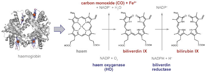
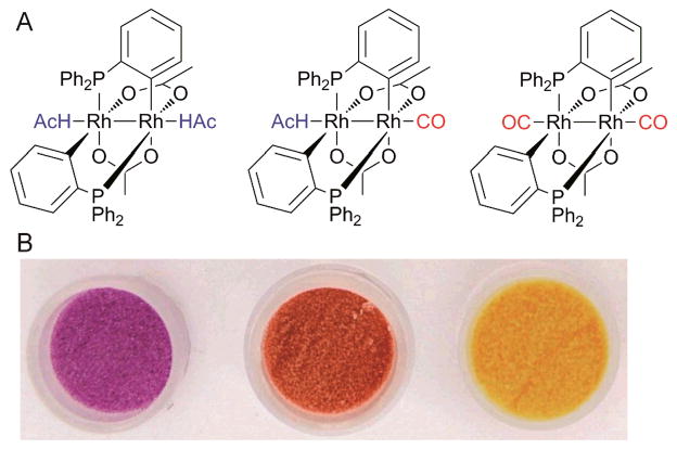
Carbon monoxide (CO) is increasingly recognized as a cell-signalling molecule akin to nitric oxide (NO). CO has attracted particular attention as a potential therapeutic agent because of its reported anti-hypertensive, anti-inflammatory and cell-protective effects. We discuss recent progress in identifying new effector systems and elucidating the mechanisms of action of CO on, e.g., ion channels, as well as the design of novel methods to monitor CO in cellular environments. We also report on recent developments in the area of CO-releasing molecules (CORMs) and materials for controlled CO application. Novel triggers for CO release, metal carbonyls and degradation mechanisms of CORMs are highlighted. In addition, potential formulations of CORMs for targeted CO release are discussed.
Figures
Similar articles
-
Carbon Monoxide and Its Controlled Release: Therapeutic Application, Detection, and Development of Carbon Monoxide Releasing Molecules (CORMs).J Med Chem. 2018 Apr 12;61(7):2611-2635. doi: 10.1021/acs.jmedchem.6b01153. Epub 2017 Sep 20. J Med Chem. 2018. PMID: 28876065 Review.
-
Design of biomaterials for intracellular delivery of carbon monoxide.Biomater Sci. 2015 Nov;3(11):1423-38. doi: 10.1039/c5bm00210a. Biomater Sci. 2015. PMID: 26252321
-
Novel lead structures and activation mechanisms for CO-releasing molecules (CORMs).Br J Pharmacol. 2015 Mar;172(6):1638-50. doi: 10.1111/bph.12688. Epub 2014 Jul 2. Br J Pharmacol. 2015. PMID: 24628281 Free PMC article. Review.
-
Development of Triggerable, Trackable, and Targetable Carbon Monoxide Releasing Molecules.Acc Chem Res. 2020 Oct 20;53(10):2273-2285. doi: 10.1021/acs.accounts.0c00402. Epub 2020 Sep 15. Acc Chem Res. 2020. PMID: 32929957 Free PMC article.
-
Metal-based carbon monoxide releasing molecules with promising cytotoxic properties.Dalton Trans. 2024 Jun 10;53(23):9612-9656. doi: 10.1039/d4dt00087k. Dalton Trans. 2024. PMID: 38808485 Review.
Cited by
-
The complex photochemistry of coumarin-3-carboxylic acid in acetonitrile and methanol.Photochem Photobiol Sci. 2022 Aug;21(8):1481-1495. doi: 10.1007/s43630-022-00238-8. Epub 2022 May 17. Photochem Photobiol Sci. 2022. PMID: 35578152
-
CO-Releasing Molecules Have Nonheme Targets in Bacteria: Transcriptomic, Mathematical Modeling and Biochemical Analyses of CORM-3 [Ru(CO)3Cl(glycinate)] Actions on a Heme-Deficient Mutant of Escherichia coli.Antioxid Redox Signal. 2015 Jul 10;23(2):148-62. doi: 10.1089/ars.2014.6151. Epub 2015 Apr 28. Antioxid Redox Signal. 2015. PMID: 25811604 Free PMC article.
-
Early processes in heme-based CO-sensing proteins.Front Mol Biosci. 2022 Nov 3;9:1046412. doi: 10.3389/fmolb.2022.1046412. eCollection 2022. Front Mol Biosci. 2022. PMID: 36406263 Free PMC article. Review.
-
Study on the Gas-Chromic Character of Pd/TiO2 for Fast Room-Temperature CO Detection.Molecules. 2024 Aug 13;29(16):3843. doi: 10.3390/molecules29163843. Molecules. 2024. PMID: 39202922 Free PMC article.
-
Simultaneous Detection of Carbon Monoxide and Viscosity Changes in Cells.Angew Chem Int Ed Engl. 2020 Nov 23;59(48):21431-21435. doi: 10.1002/anie.202008224. Epub 2020 Sep 17. Angew Chem Int Ed Engl. 2020. PMID: 32686308 Free PMC article.
References
-
- Goldbaum LR, Ramirez RG, Absalon KB. Aviat Space Environ Med. 1975;46:1289–1291. - PubMed
-
- Kim HP, Ryter SW, Choi AM. Annu Rev Pharmacol Toxicol. 2006;46:411–449. - PubMed
-
- Motterlini R, Otterbein LE. Nat Rev Drug Discov. 2010;9:728–743. - PubMed
-
- Maines MD. FASEB J. 1988;2:2557–2568. - PubMed
-
- Coburn RF. Ann N Y Acad Sci. 1970;174:11–22. - PubMed
Publication types
MeSH terms
Substances
Grants and funding
LinkOut - more resources
Full Text Sources
Other Literature Sources