Review
. 2014 Mar;63(3):819-31.
doi: 10.2337/db13-1146.

Human β-cell proliferation and intracellular signaling part 2: still driving in the dark without a road map

Affiliations
Review

Human β-cell proliferation and intracellular signaling part 2: still driving in the dark without a road map

Ernesto Bernal-Mizrachi et al. Diabetes. 2014 Mar.

Abstract

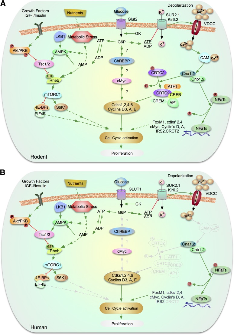
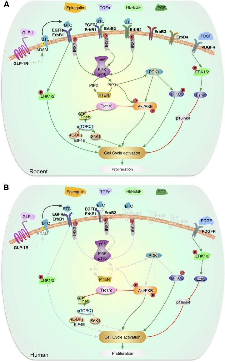
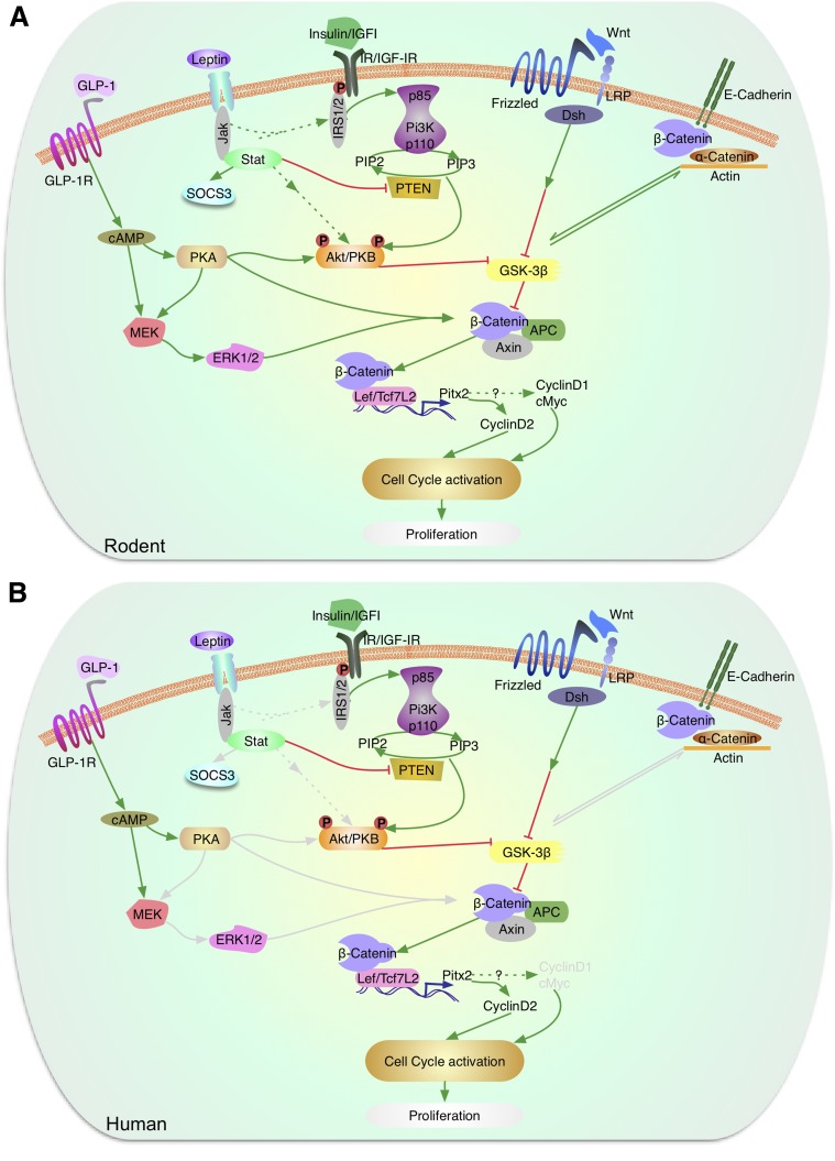
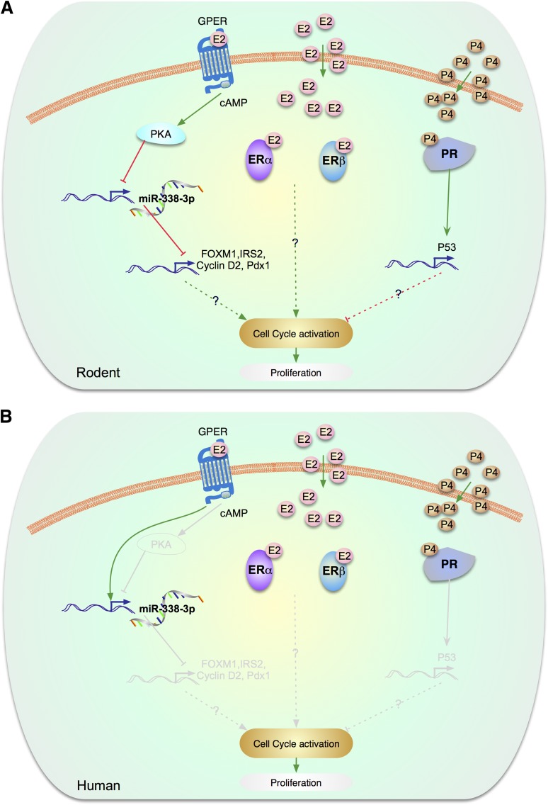
Enhancing β-cell proliferation is a major goal for type 1 and type 2 diabetes research. Unraveling the network of β-cell intracellular signaling pathways that promote β-cell replication can provide the tools to address this important task. In a previous Perspectives in Diabetes article, we discussed what was known regarding several important intracellular signaling pathways in rodent β-cells, including the insulin receptor substrate/phosphatidylinositol-3 kinase/Akt (IRS-PI3K-Akt) pathways, glycogen synthase kinase-3 (GSK3) and mammalian target of rapamycin (mTOR) S6 kinase pathways, protein kinase Cζ (PKCζ) pathways, and their downstream cell-cycle molecular targets, and contrasted that ample knowledge to the small amount of complementary data on human β-cell intracellular signaling pathways. In this Perspectives, we summarize additional important information on signaling pathways activated by nutrients, such as glucose; growth factors, such as epidermal growth factor, platelet-derived growth factor, and Wnt; and hormones, such as leptin, estrogen, and progesterone, that are linked to rodent and human β-cell proliferation. With these two Perspectives, we attempt to construct a brief summary of knowledge for β-cell researchers on mitogenic signaling pathways and to emphasize how little is known regarding intracellular events linked to human β-cell replication. This is a critical aspect in the long-term goal of expanding human β-cells for the prevention and/or cure of type 1 and type 2 diabetes.


Figures

Figure 1
Figure 1
Glucose signaling pathways to β-cell proliferation via mTOR, via ChREBP/cMyc, and via NFATs. A: Signaling mechanisms in rodent β-cells. B: Signaling molecules confirmed in human β-cells. Molecules and arrows in gray denote pathways that are known to exist in rodents, but are unstudied in human β-cells. Briefly, glucose enters the β-cell via GLUT2 (in rodents) or GLUT1 (in humans) whose kinetics ensure that phosphorylation and subsequent catabolism is proportional to blood glucose levels. Glucose is phosphorylated by GK to G-6-P and enters glycolysis. In the right side of the figure, this generates ATP, depleting ADP and AMP, which permits suppression of AMP-kinase, with resultant activation of mTOR signaling to proliferation. In parallel, in the middle of the figure, metabolism of glucose activates ChREBP, which leads to activation of cMyc and then cyclins with β-cell proliferation. In the right side of the figure, in parallel with the other pathways, glucose metabolism to generate ATP blocks potassium entry via the potassium inward rectifier/sulfonyurea receptor complex, which leads to depolarization of voltage-dependent calcium channels, and resultant calcium entry. This leads to activation of calmodulin, and thus the phosphatase, calcineurin, with resultant dephosphorylation of proliferative molecules such as the NFAT and CRTC2 families. Upon growth factor and insulin stimulation, Akt and ERK phosphorylates and inactivates TSC2, releasing the inhibition of Rheb and activation of mTOR complex 1 (mTORC1). In contrast, phosphorylation and activation of TSC2 by AMPK and GSK3β inhibits mTOR signaling. mTORC1 controls growth (cell size) and proliferation (cell number) by modulating mRNA translation through phosphorylation of 4E-BP 1, 2, and 3 and the ribosomal protein S6 kinases (S6K1 and 2). Phosphorylation of the 4E-BPs triggers their release from eIF4E and initiates cap-dependent translation. See the text for more detail. (A high-quality color representation of this figure is available in the online issue.)
Figure 2
Figure 2
Signaling by EGF and PDGF in the regulation of β-cell proliferation. Schematic representation of the signaling pathways activated by EGF family of proteins and PDGF in rodent (A) and human (B) β-cells. Multiple members of the EGF family of proteins, including BTC, EGF, HB-EGF, TGF-α, and epiregulin, have been shown to act in β-cells. ErbB family of receptors is expressed in β-cells and BTC binding to ErbB1, also called EGFR, and ErbB2 has been reported to activate the IRS2/PI3K pathway, which in turn signals via PDK-1 to modulate Akt and PKCζ. EGF binding to ErbB receptors in β-cells leads to activation of Akt and ERK signaling pathways. GLP-1 through activation of ADAM proteins can lead to the secretion of BTC from β-cells that act upon ErbB receptors. Activation of these pathways leads to enhanced rodent β-cell proliferation, an aspect unknown in human β-cells. PDGF receptors are expressed in β-cells but their expression is attenuated during aging. PDGF binding to PDGFR leads to activation of ERK1/2, increased expression of the histone methyltransferase, Ezh2, repression of the cell-cycle inhibitor p16INK4, and increased β-cell proliferation. This pathway is preserved in juvenile human β-cells and leads to enhanced replication. Gray lines are molecules and pathways that are known to exist in rodents but are unknown in human β-cells. (A high-quality color representation of this figure is available in the online issue.)
Figure 3
Figure 3
Signaling by leptin, Wnt, and β-catenin in the regulation of β-cell proliferation. A: In murine models, leptin acts via the JAK-STAT pathway to inhibit PTEN and also modulate Akt/PKB and p70S6k. Akt/PKB, which is also activated by growth factor (insulin/IGF-1) signaling, modulates GSK3β. The Wnt/frizzled pathway also regulates GSK3β, which blocks phosphorylation of β-catenin to control the expression of Lef/Tcf7L2 and cyclin D2 and potentially cyclin D1 and cMyc to activate the cell cycle and regulate proliferation. GLP-1 receptor signaling activated by GLP-1 or exendin-4 leads to elevation of cAMP and activation of protein kinase A, which can directly or indirectly via the MEK/ERK1/2 pathway phosphorylate β-catenin. Activation of the insulin or IGF-1 receptors leads to phosphorylation of serine/threonine residues in IRS2, activation of PI3K and Akt/PKB, which can, in turn, phosphorylate and inactivate GSK3β. B: In human β-cells, leptin receptors, GLP-1 receptors, and elements of the Wnt signaling pathway have been reported. However, the downstream proteins (marked in gray) that link to the proliferation response are not fully understood. (A high-quality color representation of this figure is available in the online issue.)
Figure 4
Figure 4
Estrogen/progesterone signaling pathways involved in proliferation. A: In rodent β-cells, GPER has been implicated in β-cell proliferation during pregnancy. In rodents, GPER expression is upregulated during pregnancy, which leads to a decrease in the expression of the islet microRNA, miR-338–3p, leading to increased mRNA expression of IRS-2, Pdx1, FOXM1, and cyclin D2 and stimulation of β-cell proliferation. These effects of E2/GPER are cAMP- and PKA-dependent. E2 has also been reported to increase β-cell proliferation in ovariectomized rodents with subtotal pancreatectomy. This effect was associated with an increase in the expression of IRS-2 and PDX1 proteins. B: Human β-cells. The gray lines are molecules and pathways that are known to exist in rodents but are unknown in human β-cells. The human β-cell signaling road map is underdeveloped. Although FOXM1, IRS-2, cyclin D2, PDX-1 are known to be present in human β-cell, their involvement in estrogen signaling has not been studied. Exposure to E2 reduces the level of miR-338–3p in human islet cells. However, neither E2 nor silencing of miR-338–3p elicited replication of cultured human β-cells. In addition, progesterone signaling has not been studied in human β-cells.

References

    1. Kulkarni RN, Mizrachi E-B, Ocana AG, Stewart AF. Human β-cell proliferation and intracellular signaling: driving in the dark without a road map. Diabetes 2012;61:2205–2213 - PMC - PubMed
    1. Porat S, Weinberg-Corem N, Tornovsky-Babaey S, et al. Control of pancreatic β cell regeneration by glucose metabolism. Cell Metab 2011;13:440–449 - PMC - PubMed
    1. Nakamura A, Terauchi Y, Ohyama S, et al. Impact of small-molecule glucokinase activator on glucose metabolism and beta-cell mass. Endocrinology 2009;150:1147–1154 - PubMed
    1. Guillam MT, Hümmler E, Schaerer E, et al. Early diabetes and abnormal postnatal pancreatic islet development in mice lacking Glut-2 [published correction appears in Nat Genet 1997;17:503]. Nat Genet 1997;17:327–330 - PubMed
    1. De Vos A, Heimberg H, Quartier E, et al. Human and rat beta cells differ in glucose transporter but not in glucokinase gene expression. J Clin Invest 1995;96:2489–2495 - PMC - PubMed

Publication types

MeSH terms