Review
. 2014 Aug;71(16):3027-47.
doi: 10.1007/s00018-014-1582-7. Epub 2014 Feb 21.

TPX2: of spindle assembly, DNA damage response, and cancer

Affiliations
Review

TPX2: of spindle assembly, DNA damage response, and cancer

Gernot Neumayer et al. Cell Mol Life Sci. 2014 Aug.

Abstract

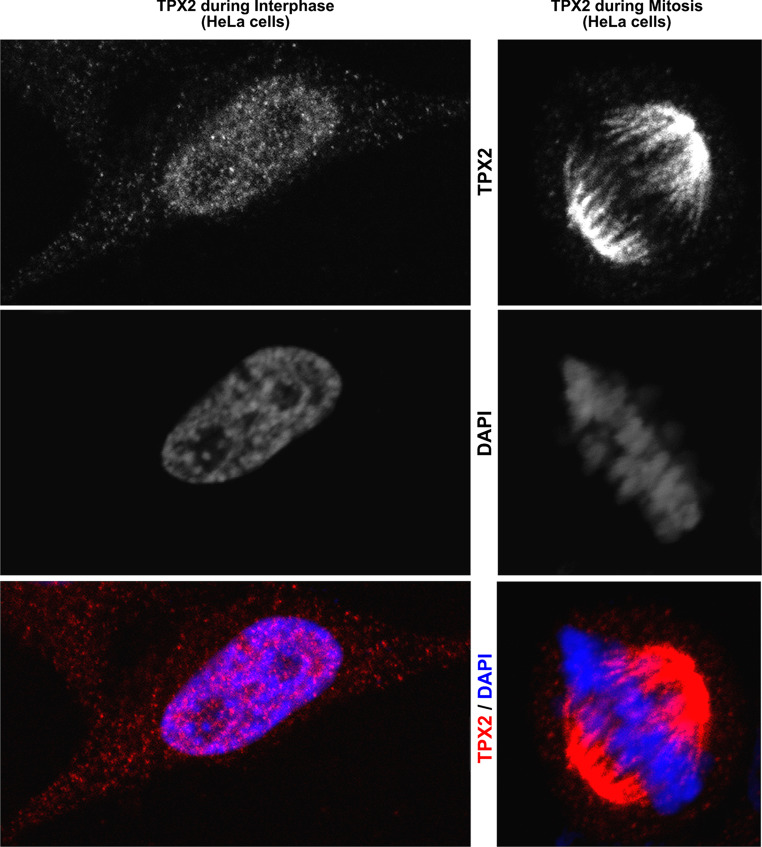
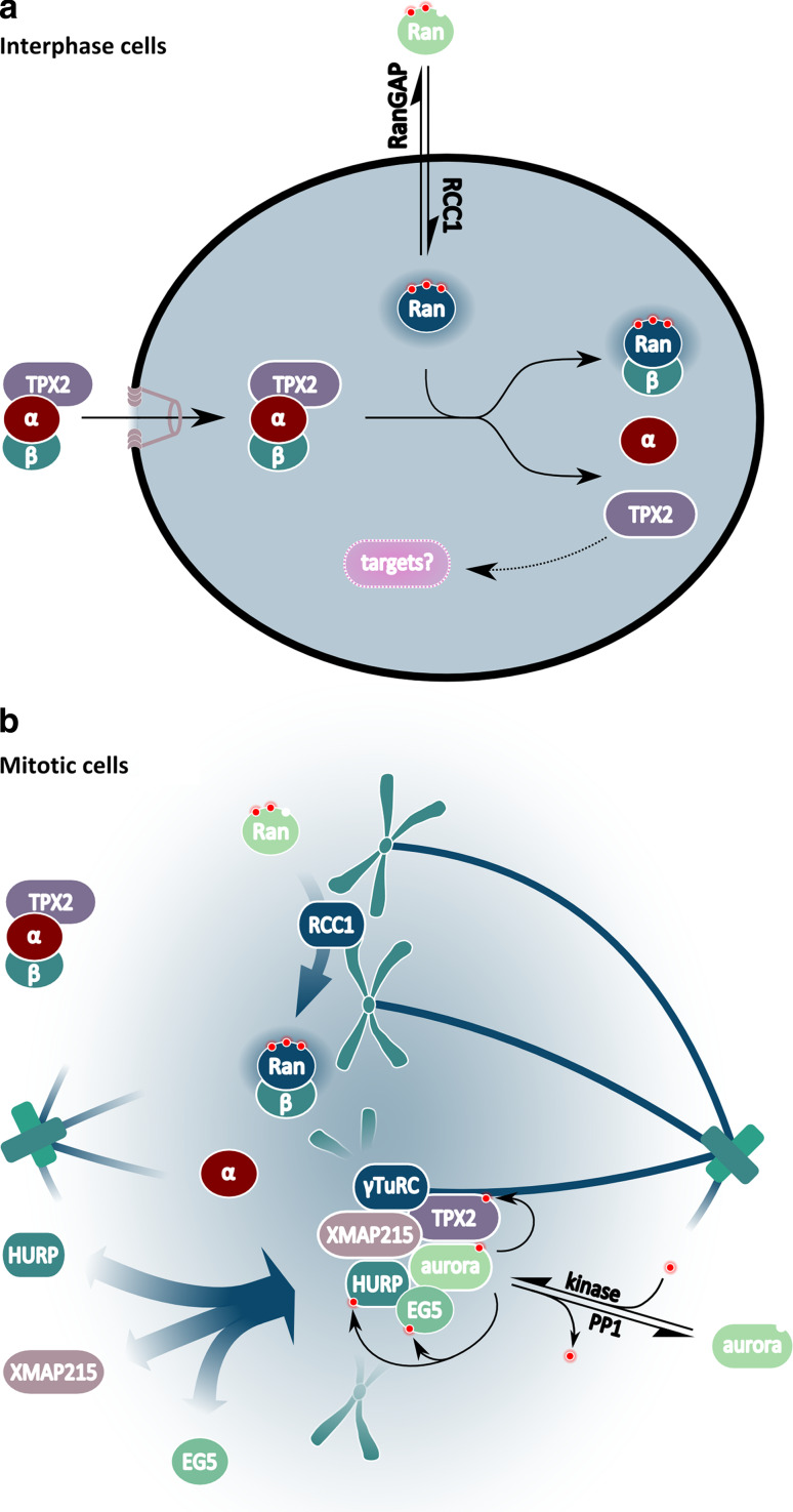
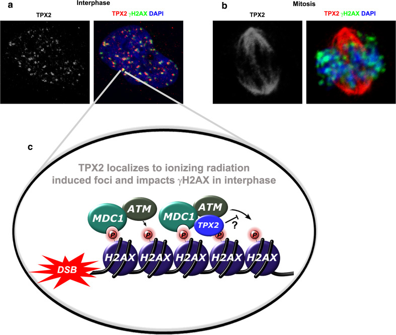
For more than 15 years, TPX2 has been studied as a factor critical for mitosis and spindle assembly. These functions of TPX2 are attributed to its Ran-regulated microtubule-associated protein properties and to its control of the Aurora A kinase. Overexpressed in cancers, TPX2 is being established as marker for the diagnosis and prognosis of malignancies. During interphase, TPX2 resides preferentially in the nucleus where its function had remained elusive until recently. The latest finding that TPX2 plays a role in amplification of the DNA damage response, combined with the characterization of TPX2 knockout mice, open new perspectives to understand the biology of this protein. This review provides an historic overview of the discovery of TPX2 and summarizes its cytoskeletal and signaling roles with relevance to cancer therapies. Finally, the review aims to reconcile discrepancies between the experimental and pathological effects of TPX2 overexpression and advances new roles for compartmentalized TPX2.


Figures

Fig. 1
Fig. 1
The localization of TPX2 during interphase and mitosis. A cell cycle time of ~24 h, with ~1 h for mitosis, appears to be standard for several human cell lines (e.g., HeLa cells) in culture [2, 185]. During interphase, taking up the majority of the cell cycle time, TPX2 is predominantly a nuclear protein (left). During mitosis (right), TPX2 strongly associates with MTs of the spindle. Confocal images of HeLa cells with endogenous TPX2 (red/white) are shown. DNA was visualized with DAPI (blue/white)
Fig. 2
Fig. 2
The MAP functions of TPX2. a Without TPX2, nucleation of MTs is inefficient and Xklp2 localizes to the plus ends of MTs. b MT nucleation is enhanced by TPX2. TPX2 also targets Xklp2 to MT minus ends. The latter function is dependent on the dynein/dynactin molecular motors (not shown). c TPX2 acts in concert with Augmin to form branch points that nucleate new MTs at a low-angle, allowing amplification of MTs mass. In addition, TPX2 bundles and organizes polymerized MTs. d In brief, TPX2 regulates the formation of the mitotic spindle via several MTs-based mechanisms: (1) TPX2 localizes Xklp2 to the spindle poles, which is important for spindle bipolarity, (2) TPX2 mediates the nucleation and bundling of MTs, and (3) TPX2 is required for the formation of low-angle junctions that nucleate new MTs
Fig. 3
Fig. 3
Compartmentalized functions of TPX2. a During interphase, TPX2 binds directly to cytoplasmic Importinα, which binds to Importinβ. The resulting TPX2-Importinα/β complex then shuttles into the nucleus. Directed transport is enabled by a low cytoplasmic concentration of RanGTP that is maintained by the cytoplasmic factors RanGAP and RanBP1+2 (not shown). A high nuclear concentration of RanGTP, maintained by the chromatin bound RanGEF RCC1, causes the dissociation of the TPX2-Importinα/β complex. Specifically, after RanGTP binding to Importinβ, the affinity of Importinα for TPX2 decreases, thereby leading to the dissociation of the entire complex and the release of TPX2. In the nucleus, TPX2 participates in DNA damage response via unknown molecular mechanisms (not shown; see Fig. 4). b During mitosis, a high concentration of RanGTP (blue gradient shading) is found in the vicinity of the chromosomes that are associated with RCC1. RanGTP causes the release of TPX2 from nuclear import receptors (see a for details). Importin-free TPX2 nucleates MTs (not shown; see Fig. 2), activates a sub-population of the Aurora A kinase (via direct binding and protection from protein phosphatase 1; PP1) and mediates the association of Aurora A with spindle MTs. Aurora A kinase activity is required for formation of the EXTAH complex that participates in spindle morphogenesis. Note that TPX2 bound by Importinα (in areas of low RanGTP concentrations) cannot activate or associate with Aurora A. Numerous other modes of Aurora A activation have also been identified (not shown). Phosphate groups are depicted as small red filled circles. See text for details
Fig. 4
Fig. 4
A novel and first nuclear function for TPX2. a During DNA damage response elicited by ionizing radiation in interphase U2OS cells, TPX2 co-localizes with γ-H2AX at chromosomal breakage sites. The shown ionizing radiation-induced foci [i.e. white/red (TPX2) or green (γ-H2AX) dots] demonstrate accumulation of γ-H2AX and TPX2 at DNA double-strand breaks. b In mitotic U2OS cells undergoing intrinsically hampered DNA damage response, TPX2 is localized to the mitotic spindle and excluded from the γ-H2AX-positive ionizing radiation-induced foci. DNA was visualized with DAPI. c Model for TPX2’s function during DNA damage response: MDC1 can simultaneously bind γ-H2AX and the ATM kinase. The MDC1-recruited ATM generates additional γ-H2AX. TPX2 localizes to γ-H2AX, MDC1, and ATM positive ionizing radiation-induced foci and inhibits MDC1/ATM-mediated phosphorylation of H2AX via unknown mechanisms (see text for details). DSB DNA double-strand break

References

    1. Heidebrecht HJ, Buck F, Steinmann J, Sprenger R, Wacker HH, Parwaresch R. p100: a novel proliferation-associated nuclear protein specifically restricted to cell cycle phases S, G2, and M. Blood. 1997;90(1):226–233. - PubMed
    1. Gruss OJ, Wittmann M, Yokoyama H, Pepperkok R, Kufer T, Sillje H, Karsenti E, Mattaj IW, Vernos I. Chromosome-induced microtubule assembly mediated by TPX2 is required for spindle formation in HeLa cells. Nat Cell Biol. 2002;4(11):871–879. - PubMed
    1. Wittmann T, Boleti H, Antony C, Karsenti E, Vernos I. Localization of the kinesin-like protein Xklp2 to spindle poles requires a leucine zipper, a microtubule-associated protein, and dynein. J Cell Biol. 1998;143(3):673–685. - PMC - PubMed
    1. Vernos I, Heasman J, Wylie C. Multiple kinesin-like transcripts in Xenopus oocytes. Dev Biol. 1993;157(1):232–239. - PubMed
    1. Boleti H, Karsenti E, Vernos I. Xklp2, a novel Xenopus centrosomal kinesin-like protein required for centrosome separation during mitosis. Cell. 1996;84(1):49–59. - PubMed

Publication types

MeSH terms

LinkOut - more resources