. 2014;6(4):426-34.
doi: 10.1159/000358834. Epub 2014 Feb 15.

Relative contribution of c1q and apoptotic cell-surface calreticulin to macrophage phagocytosis

Affiliations

Relative contribution of c1q and apoptotic cell-surface calreticulin to macrophage phagocytosis

Mélanie Verneret et al. J Innate Immun. 2014.

Abstract

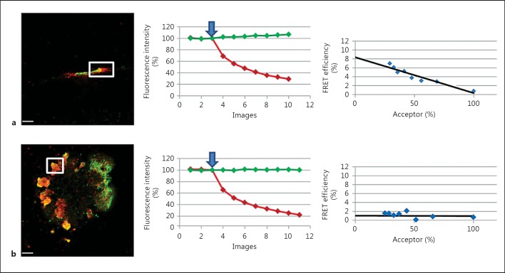
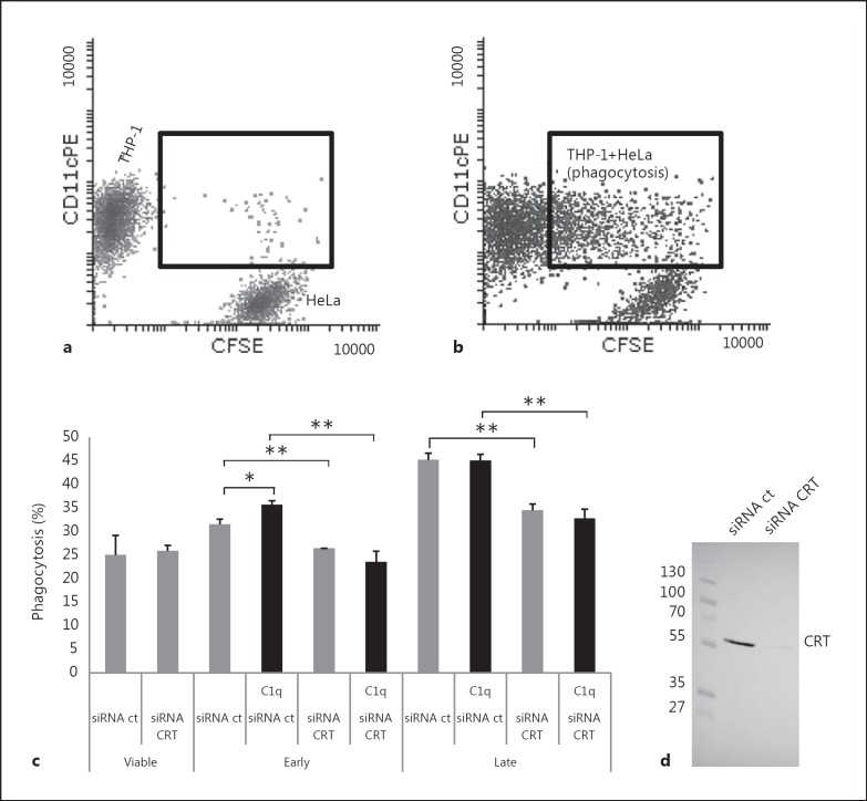
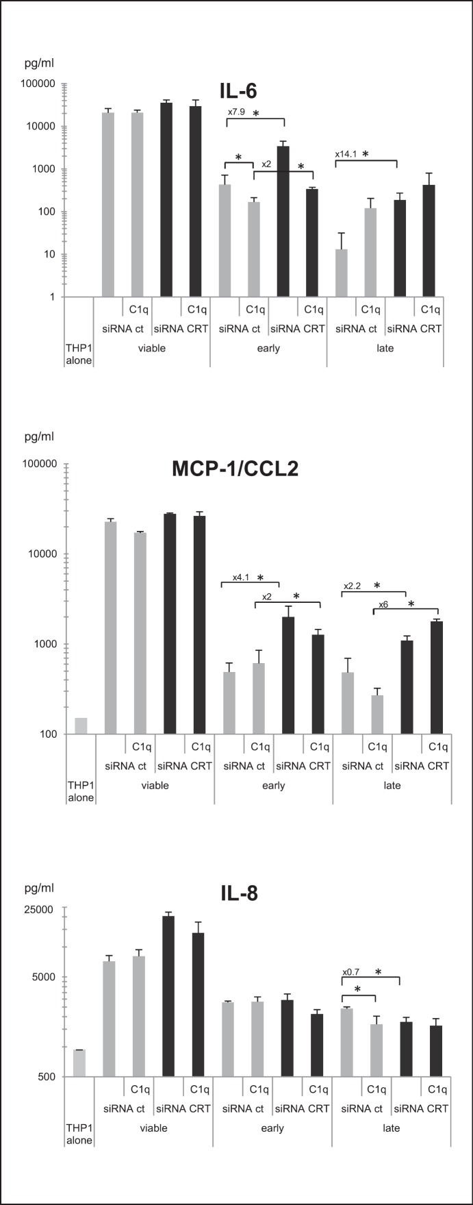
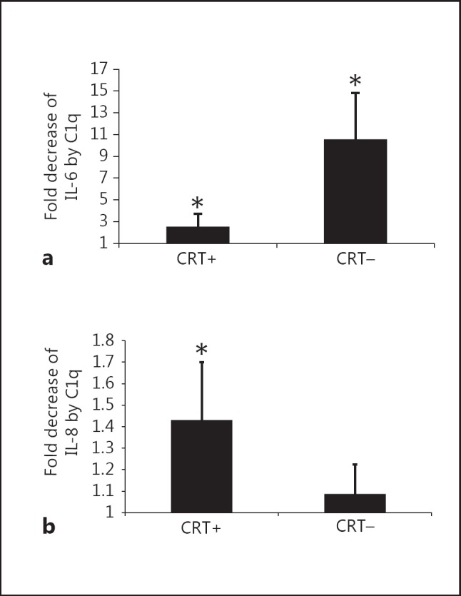
C1q has been shown to recognize apoptotic cells, to enhance their uptake and to modulate cytokine release by phagocytes and thus promote immune tolerance. Surface-exposed calreticulin (CRT), known as a C1q receptor, is also considered to be an early eat-me signal that enhances the phagocytosis of apoptotic cells and is capable of eliciting an immunogenic response. However, the molecular mechanisms that trigger these functions are not clear. We hypothesized that CRT and C1q might act together in these processes. We first showed, by means of fluorescence resonance energy transfer (FRET), that CRT interacts with the C1q globular region at the surface of early apoptotic cells. Next, we pointed out that knockdown of CRT on early apoptotic HeLa cells impairs the enhancement effect of C1q on their uptake by THP-1 monocyte-derived macrophages. Furthermore, a deficiency of CRT induces contrasting effects on cytokine release by THP-1 macrophages, increasing interleukin (IL)-6 and monocyte chemotactic protein 1/CCL2 and decreasing IL-8. Remarkably, these effects were greatly reduced when apoptotic cells were opsonized by C1q, which counterbalanced the effect of the CRT deficiency. These results demonstrate that CRT-C1q interaction is involved in the C1q bridging function and they highlight the particular ability of C1q to control the phagocyte inflammatory status, i.e. by integrating the molecular changes that could occur at the surface of dying cells.


Figures

Fig. 1
Fig. 1
FRET study of C1qGR and CRT at the surface of apoptotic HeLa cells. FRET efficiency was estimated by photobleaching of the acceptor dye (Cy-3) on nonpermeabilized cells incubated with C1qGR and immunolabeled for CRT (Alexa-488) and C1qGR (Cy-3) as described in Materials and Methods. a Apoptotic cell without visible membrane bleb. b Apoptotic cell with characteristic membrane blebs. Regions used for the acceptor photobleaching and FRET analysis are shown. Bars: 5 µm. Middle: curves corresponding to the normalized fluorescence intensities of both dyes (Cy-3 in red and Alexa-488 in green) expressed as a percent of the signal measured before the gradual photobleaching started (blue arrow). Right: FRET efficiency (percent of acceptor fluorescence intensity increase) is expressed as a function of the percent of the normalized acceptor fluorescence intensity.
Fig. 2
Fig. 2
Effect of CRT deficiency and of exogenous C1q on the uptake of early and late apoptotic cells. Early or late apoptotic HeLa cells labeled with CFSE, opsonized or not with C1q (25 µg/ml), were incubated with PMA-treated THP-1 cells at a ratio of 1:1 for 1 h at 37°C. THP-1 macrophages were labeled with an anti-CD11c-PE antibody just prior to flow cytometry analysis. a Dot-plot of a negative control phagocytosis performed at 4°C (negative control). b Dot-plot of the phagocytosis at 37°C (corresponding to late apoptotic CRT-deficient cells). a, b Inset shows macrophages that had engulfed HeLa cells (double-labeled cells). c Phagocytosis is expressed as the percent of the double-labeled cells in the macrophage population (i.e. CD11c-positive cells). Data are the mean ± SD of triplicates of a representative experiment of 5. * p ≤ 0.05, ** p ≤ 0.005, ANOVA 1-way test. d Representative SDS-PAGE analysis and immunoblotting of CRT expressed on siRNA-treated HeLa cells; 20 µg of soluble proteins were analyzed. The molecular mass markers (expressed in kilodaltons) are shown.
Fig. 3
Fig. 3
Cytokines released from THP-1 macrophages during the uptake of CRT-deficient apoptotic cells. Cytokine production of PMA-treated THP-1 monocytes was analyzed using BDBiosciences Cytometric BeadArray in the supernatant of macrophages fed with early or late apoptotic cells deficient or not for CRT (i.e. siRNA CRT or siRNA ct cells). When indicated, early or late apoptotic cells were opsonized by C1q. Supernatants were collected 18 h after the phagocytosis assay. Data are the average concentrations ± SD from measurements of 3 independent experiments. * p ≤ 0.05, Student t test. Control cytokine levels produced by THP-1 alone or fed with viable cells are shown. Fold modulation of the cytokines released in the siRNA CRT versus siRNA ct conditions is reported when differences were statistically significant.
Fig. 4
Fig. 4
C1q modulation of IL-6 and IL-8 released from macrophages during the uptake of apoptotic cells. Cytokine levels were measured by analysis of the supernatant of differentiated THP-1 macrophages and fed with early (IL-6) or late (IL-8) apoptotic cells as described for figure 3. Selected results of figure 3 are expressed as fold difference in cytokine concentration (a IL-6 and b IL-8) compared to control levels from phagocytes, which had ingested apoptotic cells in the absence of C1q. Data are plotted as average fold decreases ± SD from 3 experiments. * p ≤ 0.05 (C1q-treated levels vs. control levels in the absence of C1q, Student t test). CRT+ = siRNA control-treated cells; CRT- = siRNA CRT-treated cells.

References

    1. Gaboriaud C, Frachet P, Thielens NM, Arlaud GJ. The human C1q globular domain: structure and recognition of non-immune self ligands. Front Immunol. 2011;2:92. - PMC - PubMed
    1. Korb LC, Ahearn JM. C1q binds directly and specifically to surface blebs of apoptotic human keratinocytes: complement deficiency and systemic lupus erythematosus revisited. J Immunol. 1997;158:4525–4528. - PubMed
    1. Navratil JS, Watkins SC, Wisnieski JJ, Ahearn JM. The globular heads of C1q specifically recognize surface blebs of apoptotic vascular endothelial cells. J Immunol. 2001;166:3231–3239. - PubMed
    1. Nayak A, Ferluga J, Tsolaki AG, Kishore U. The non-classical functions of the classical complement pathway recognition subcomponent C1q. Immunol Lett. 2010;131:139–150. - PubMed
    1. Fraser DA, Laust AK, Nelson EL, Tenner AJ. C1q differentially modulates phagocytosis and cytokine responses during ingestion of apoptotic cells by human monocytes, macrophages, and dendritic cells. J Immunol. 2009;183:6175–6185. - PMC - PubMed

Publication types