. 2014 Mar 14;343(6176):1221-8.
doi: 10.1126/science.1243462. Epub 2014 Feb 20.

Specific and nonhepatotoxic degradation of nuclear hepatitis B virus cccDNA

Affiliations

Specific and nonhepatotoxic degradation of nuclear hepatitis B virus cccDNA

Julie Lucifora et al. Science. .

Abstract

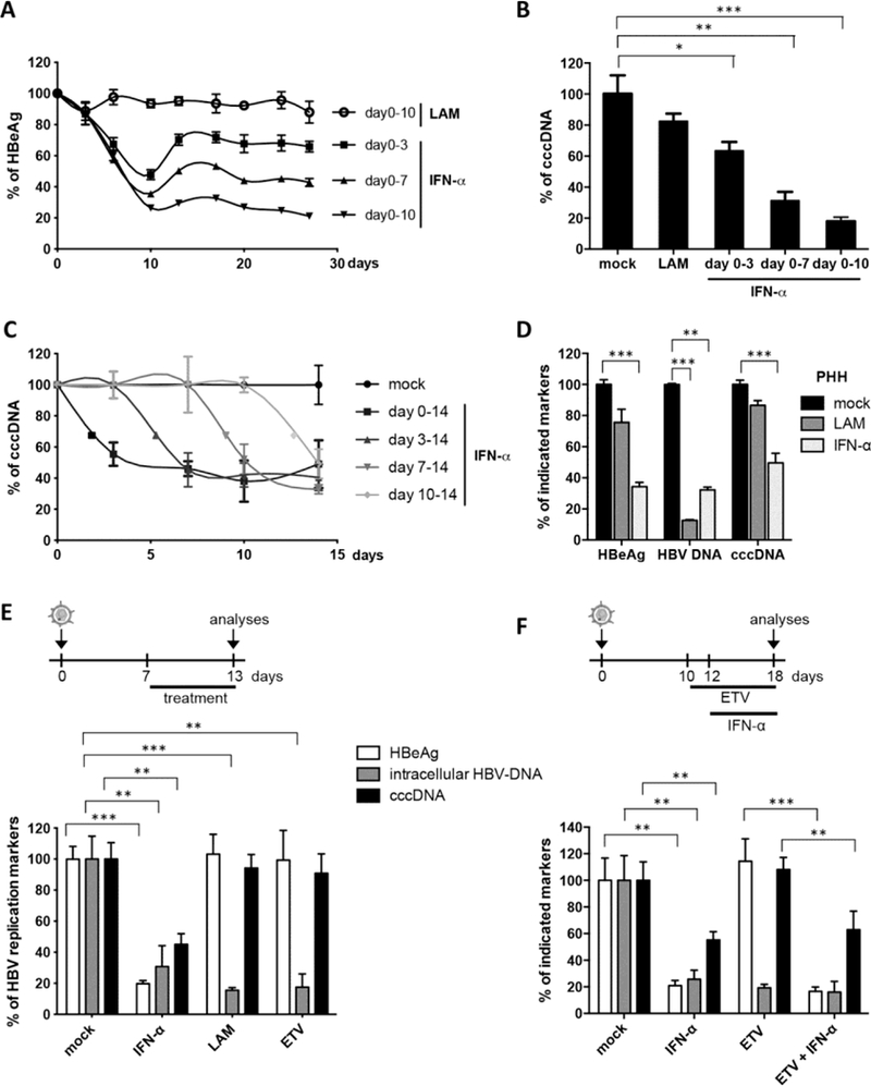
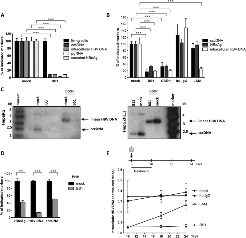
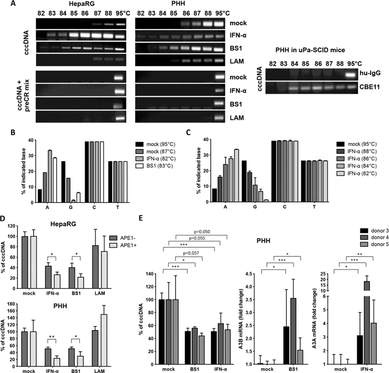
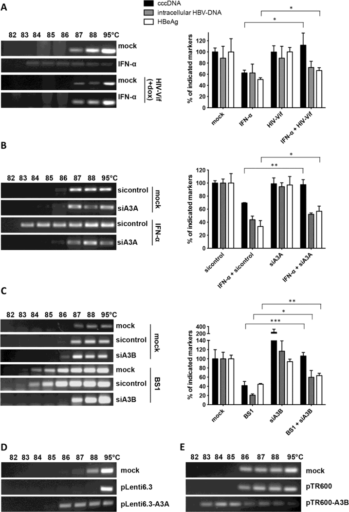
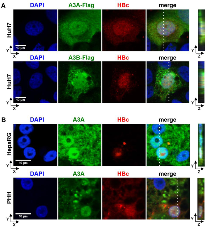
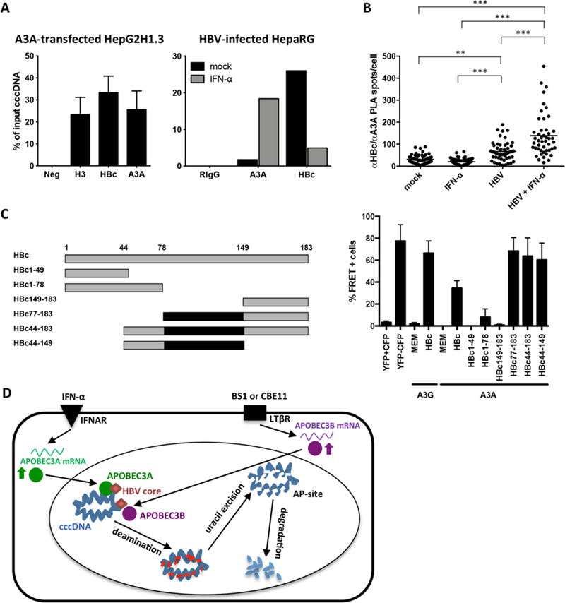
Current antiviral agents can control but not eliminate hepatitis B virus (HBV), because HBV establishes a stable nuclear covalently closed circular DNA (cccDNA). Interferon-α treatment can clear HBV but is limited by systemic side effects. We describe how interferon-α can induce specific degradation of the nuclear viral DNA without hepatotoxicity and propose lymphotoxin-β receptor activation as a therapeutic alternative. Interferon-α and lymphotoxin-β receptor activation up-regulated APOBEC3A and APOBEC3B cytidine deaminases, respectively, in HBV-infected cells, primary hepatocytes, and human liver needle biopsies. HBV core protein mediated the interaction with nuclear cccDNA, resulting in cytidine deamination, apurinic/apyrimidinic site formation, and finally cccDNA degradation that prevented HBV reactivation. Genomic DNA was not affected. Thus, inducing nuclear deaminases-for example, by lymphotoxin-β receptor activation-allows the development of new therapeutics that, in combination with existing antivirals, may cure hepatitis B.


Figures

Fig. 1.
Fig. 1.. Degradation of cccDNA in IFN-α treated HepaRG cells and primary human hepatocytes.
(A, B, C, E, and F) HBV-infected dHepaRG were treated with IFN-α at day 10 post-infection (dpi). Different regimens of treatment were applied as indicated. (D) HBVinfected primary human hepatocyte (PHH) were treated with IFN-α at dpi 3 for 13 days. Levels of HBeAg, total intracellular DNA and cccDNA are given relative to mock treated cells. LAM: lamivudine; ETV: entecavir. Mean values +/− standard deviation of replicates from independent experiments are given; data were analyzed by t test. * p < 0.05, ** p < 0.01 and *** p < 0.001.
Fig. 2.
Fig. 2.. LTβR-activation inhibits HBV infection and leads to cccDNA degradation in HepaRG cells and PHH.
(A and B) HBV-infected dHepaRG were treated with BS1, CBE11, hu-IgG control or lamivudine (LAM). (A) Treatment started 24h before infection for 12 days or (B) at 18 dpi for 10 days. Levels of the indicated HBV markers as well as cell viability are given relative to untreated controls (mock). (C) cccDNA levels were analyzed after 14 days of BS1 treatment by Southern blot in HBV-infected dHepaRG and HBV-replicating HepG2H1.3 cells. Supercoiled cccDNA bands were identified by their expected size and linearization upon EcoRI digestion (3,2 kb). (D) PHH were infected with HBV and treated with BS1 at 7 dpi for 10 days. Levels of the indicated HBV markers were compared to untreated PHH of the same donor (donor 3) (mock). (E) HBV-infected dHepaRG were treated with BS1, hu-IgG control or LAM. Intracellular HBV-DNA was analyzed 8 and 14 days after treatment cessation. Mean values +/− standard deviation of replicates from independent experiments are given; data were analyzed by t test. * p < 0.05, *** p < 0.001.
Fig. 3.
Fig. 3.. Deamination and AP-site formation in cccDNA upon IFN-α treatment and LTβR-activation.
(A) dHepaRG (left) and PHH (middle panel) were infected with HBV and treated with IFN-α, BS1 or LAM. Human chimeric uPA/SCID mice were treated with CBE11 or hu-IgG control (right panel). 3D-PCR analyses were performed on cccDNA left either untreated (upper panels) or treated with a PreCR mix (lower panels). (B and C) 3D-PCR products from HBV-infected dHepaRG cells treated as indicated (IFN-α, BS1 or mock) were cloned and sequenced and mutations were analyzed. (D) Total DNA extracts from HBV-infected cells treated as indicated were digested with APE1, and cccDNA content was compared to mock-treated cells. In (B), (C), and (D), mean values +/− standard deviation of biological triplicates from two independent experiments are given; data were analyzed by t test. * p < 0.05, ** p < 0.01. (E) PHH were infected with HBV and treated with BS1 or IFN-α at 7 dpi for 10 days. Levels of the indicated cccDNA as well as A3A and A3B mRNA expression were compared to untreated PHH (mock) of the same donor.
Fig. 4.
Fig. 4.. Analysis of cccDNA deamination and degradation.
(A to C) cccDNA denaturation was analyzed by 3D-PCR (left panels); levels of HBeAg, total intracellular DNA and cccDNA are given relative to mock treated cells (right panels). (A) dHepaRG-tA-Vif cells treated with IFN-α for 10 days with and without doxycycline (dox)-induced HIV-Vif expression. HBV-infected dHepaRG cells treated with (B) IFN-α or (C) BS1 transfected with siRNA against A3A or A3B, respectively, or sequence nonspecific siRNA (sicontrol). Mean values +/− standard deviation of independent replicates and experiments are given; data were analyzed by t test. * p < 0.05, ** p < 0.01 and *** p < 0.001. (D) cccDNA denaturation analysis by 3D-PCR in HepG2-H1.3 cells overexpressing A3A or (E) A3B from lentiviral vector plasmid pLenti6.3 or pTR600, respectively, for 5 days.
Fig. 5.
Fig. 5.. Co-localization of A3A and A3B with HBV core protein (HBc).
(A) HuH7 cells were co-transfected with an HBV1.1-fold genome and A3A-Flag or A3B-Flag expressing plasmids and stained using DAPI, anti-HBc and anti-FLAG antibodies. (B) HBV-infected dHepaRG and PHH were treated with IFN-α at day 7 post infection for 3 days. A3A and HBc were analyzed by immunofluorescence staining. Right panels indicate z stacks taken at the dotted lines.
Fig. 6.
Fig. 6.. Interaction of A3A, HBV core protein (HBc) and cccDNA.
(A) Chromatin immunoprecipitation (ChIP) was performed using lysates of HepG2H1.3 cells transfected with A3Aexpressing plasmid, or HBV-infected dHepaRG cells treated with IFN-α for 3 days. IPs using antibodies against histone H3, A3A, HBc and control rabbit IgG (RIgG) were analyzed by qPCR for cccDNA. (B) Interaction between HBc and A3A was assessed by proximity ligation assay (PLA) in HBV-infected, IFN-α treated dHepaRG. PLA-spots were quantified in single cells by software-based spot-counting. Data were analyzed by one-way ANOVA. ** p < 0.01 and *** p < 0.001. (C) Serial HBV core-deletion mutants (left panel) were fused to CFP and interaction with A3A-YFP was assessed by FACS-FRET in HuH7.5 hepatoma cells (right panel). Cells cotransfected with CFP and YFP served as controls to exclude false positive FRET and subtract background signals. A CFP-YFP fusion construct was used as positive control. Mean values ± standard deviation of FRET-positive cells from 3–4 independent experiments are given. Black boxes indicate shared regions of HBc mutants giving a FRET signal. (D) Model of cccDNA degradation induced by IFN-α treatment or LTβR-activation.

Comment in

References

    1. Protzer U, Maini MK, Knolle PA, Living in the liver: hepatic infections. Nat. Rev. Immunol 12, 201–213 (2012). doi:10.1038/nri3169 Medline - DOI - PubMed
    1. Zoulim F, Hepatitis B virus resistance to antiviral drugs: where are we going? Liver Int 31, (Suppl 1), 111–116 (2011). doi:10.1111/j.1478-3231.2010.02399.x Medline - DOI - PMC - PubMed
    1. Wursthorn K, Lutgehetmann M, Dandri M, Volz T, Buggisch P, Zollner B, Longerich T, Schirmacher P, Metzler F, Zankel M, Fischer C, Currie G, Brosgart C, Petersen J, Peginterferon alpha-2b plus adefovir induce strong cccDNA decline and HBsAg reduction in patients with chronic hepatitis B. Hepatology 44, 675–684 (2006). doi:10.1002/hep.21282 Medline - DOI - PubMed
    1. Guidotti LG, Ando K, Hobbs MV, Ishikawa T, Runkel L, Schreiber RD, Chisari FV, Cytotoxic T lymphocytes inhibit hepatitis B virus gene expression by a noncytolytic mechanism in transgenic mice. Proc. Natl. Acad. Sci. U.S.A 91, 3764–3768 (1994). doi:10.1073/pnas.91.9.3764 Medline - DOI - PMC - PubMed
    1. Guidotti LG, Rochford R, Chung J, Shapiro M, Purcell R, Chisari FV, Viral clearance without destruction of infected cells during acute HBV infection. Science 284, 825–829 (1999). doi:10.1126/science.284.5415.825 Medline - DOI - PubMed

Publication types

MeSH terms

Associated data