. 2014 Sep;16(9):1196-209.
doi: 10.1093/neuonc/nou020. Epub 2014 Feb 20.

Proteomic screening identifies a YAP-driven signaling network linked to tumor cell proliferation in human schwannomas

Affiliations

Proteomic screening identifies a YAP-driven signaling network linked to tumor cell proliferation in human schwannomas

Alizée Boin et al. Neuro Oncol. 2014 Sep.

Abstract

Background: Inactivation of the NF2 gene predisposes to neurofibromatosis type II and the development of schwannomas. In vitro studies have shown that loss of NF2 leads to the induction of mitogenic signaling mediated by receptor tyrosine kinases (RTKs), MAP kinase, AKT, or Hippo pathways. The goal of our study was to evaluate the expression and activity of these signaling pathways in human schwannomas in order to identify new potential therapeutic targets.

Methods: Large sets of human schwannomas, totaling 68 tumors, were analyzed using complementary proteomic approaches. RTK arrays identified the most frequently activated RTKs. The correlation between the expression and activity of signaling pathways and proliferation of tumor cells using Ki67 marker was investigated by reverse-phase protein array (RRPA). Finally, immunohistochemistry was used to evaluate the expression pattern of signaling effectors in the tumors.

Results: We showed that Her2, Her3, PDGFRß, Axl, and Tie2 are frequently activated in the tumors. Furthermore, RRPA demonstrated that Ki67 levels are linked to YAP, p-Her3, and PDGFRß expression levels. In addition, Her2, Her3, and PDGFRß are transcriptional targets of Yes-associated protein (YAP) in schwannoma cells in culture. Finally, we observed that the expression of these signaling effectors is very variable between tumors.

Conclusions: Tumor cell proliferation in human schwannomas is linked to a signaling network controlled by the Hippo effector YAP. Her2, Her3, PDGFRß, Axl, and Tie2, as well as YAP, represent potentially valuable therapeutic targets. However, the variability of their expression between tumors may result in strong differences in the response to targeted therapy.

Keywords: Neurofibromatosis type 2; YAP; proteomic; schwannoma; signaling.


Figures

Fig. 1.
Fig. 1.
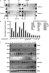
Only a limited set of receptor tyrosine kinases (RTKs) are frequently activated in human schwannomas. (A) Four examples of RTK arrays membrane incubated with extracts from human vestibular schwannomas. RTKs were considered activated when the signal was more intense than the control immunoglobulin spots. Strong signals at each corner correspond to a phosphotyrosine positive control. (B) Quantification of the percentage of tumors expressing specific activated RTKs. Fifteen out of 42 RTKs were detected in their activated form. The active RTKs that were not detected are also indicated. (C) Expression of EGFR, Her2, Her3, Axl, PDGFRß, and Tie2 were confirmed by Western blot in a series of 12 human schwannoma extracts. Extracts from HEI193 human schwannoma cell line (SC) were used for comparison. Actin was used as a loading control.
Fig. 2.
Fig. 2.
Immunohistochemical evaluation of receptor tyrosine kinase expression in human schwannomas. The expression of Her2, Her3, Axl, PDGFRß, and EGFR was evaluated by immunohistochemistry on schwannoma sections. The percentage of positive tumors is indicated on the right. No EGFR was detected in schwannomas, although the antibody gave a clear signal in the control placenta section (insert).
Fig. 3.
Fig. 3.
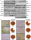
Major mitogenic signaling pathways are active in human schwannomas. (A) The expression and phosphorylation of major signaling pathways known to be regulated by merlin were evaluated by Western blot in a series of 12 human schwannomas. HEI193 extracts were used for comparison. Actin was used as a loading control. (B) Immunohistochemistry was used to evaluate the expression of phosphorylated Akt473, GSK3ß9, and MAPK202/204 as well as YAP, STAT3, and ß-catenin in human schwannoma sections. The enlarged insert for ß-catenin clearly shows negative nuclear staining. The percentage of tumor with a positive detection of the markers is indicated.
Fig. 4
Fig. 4
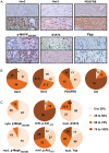
Schwannomas are very heterogeneous for the percentage of cells expressing signaling effectors. (A) The heterogeneity of the expression of Her2, Her3, PDGFRß, phospho-MAPK202/204, STAT3, and YAP was evaluated by immunohistochemistry. The top panel provides an example of a low percentage of positive cells, whereas the lower panel represents tumors with a high percentage of labeled cells. (B) The graph represents the percentage of tumors that express Her2, Her3, PDGFRß, or Axl in <25%, between 25% and 50%, between 50% and 75%, and in more than 75% of cells. (C) The same experiment was performed for the activated forms of MAPK, Akt, STAT3, and YAP. The phosphorylated forms of MAPK and Akt were independently evaluated in the cytoplasm and in the nucleus.
Fig. 5.
Fig. 5.
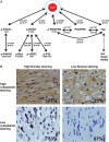
Reverse phase protein array analysis of human schwannomas reveals signaling network correlated with proliferation marker ki67. (A) The expression of YAP, PDGFRß, phospho-PDGFRß1021, phospho-Her31289, and Phospho-STAT3705 are correlated with the expression of Ki67 in human schwannoma samples. The P value and correlation coefficient are indicated for each correlation. For these 5 markers, the other significant correlations are indicated. (B) YAP intensity in the nucleus and the cytoplasm were scored by immunohistochemistry. High nuclear staining was primarily associated with high cytoplasmic staining and vice versa. The percentage of tumors represented in each case is provided. A Spearman correlation test indicates that the intensities in the nucleus and the cytoplasm are linked (P = .0009), suggesting that the activity of YAP is a function of its expression level.
Fig. 6.
Fig. 6.
The most frequently activated receptor tyrosine kinases in schwannomas are transcriptional targets of YAP. (A) A HEI193 human schwannoma cell line was created that stably overexpresses GFP-YAP contructs. The fusion protein accumulates in the nucleus (left). Western blot confirms the overexpression of GFP-YAP in the cells. (B) Western blot shows that the expression of Her2, Her3, PDGFRß, and survivin is markedly induced upon GFP-YAP expression. Axl levels appear essentially unaffected. (C) Quantitative Real Time-PCR confirms that the induction of Her2, Her3, and PDGFRß by GFP-YAP is of transcriptional orgin. Survivin, a known target gene of YAP, was used as a positive control. On the contrary, Axl is not induced by YAP. The average induction folds are indicated. The graph is representative of 6 experiments. (D) HEI193 cells overexpressing GFP-YAP treated with the YAP/Tead dissociating compound verteporfin show a strong decrease in Tead-mediated transcriptional activity measured by luciferase reporter assay. (E) The protein levels of Her3, PDGFRß, and survivin decrease in response to treatment with verteporfin. (F) Similarly, quantitative Real Time-PCR indicates that verteporfin treatment of HEI193 GFP-YAP cells reduces mRNA levels of survivin, Her3, and PDGFRß. Her2 expression is unaffected. (* = P value <.05; N/S = not significant)
Fig. 7.
Fig. 7.
Model of signaling pathway regulation in human schwannomas. When expressed in Schwann cells, merlin inhibits growth factor receptor delivery at the plasma membrane, as we previously demonstrated (1). Merlin also blocks YAP nuclear accumulation (2) leading to the inhibition of proliferation (3). In schwannomas, where merlin expression is lost (4), YAP accumulates in the nucleus (5) and stimulates the expression of PDGFRß, Her2, Her3, and possibly Axl as well as some of their ligands such as PDGF and Gas6 (6). In the absence of merlin, these receptors accumulate at the cell surface (7) where they stimulate mitogenic signaling pathway (8) proliferation and survival (9). In parallel, Tie2 expression may stimulate the development of new blood vessels (10) contributing to tumor development.

References

    1. Asthagiri AR, Parry DM, Butman JA, et al. Neurofibromatosis type 2. Lancet. 2009;373(9679):1974–1986. - PMC - PubMed
    1. Rouleau GA, Merel P, Lutchman M, et al. Alteration in a new gene encoding a putative membrane-organizing protein causes neuro-fibromatosis type 2. Nature. 1993;363(6429):515–521. - PubMed
    1. Morrison H, Sperka T, Manent J, et al. Merlin/neurofibromatosis type 2 suppresses growth by inhibiting the activation of Ras and Rac. Cancer Res. 2007;67(2):520–527. - PubMed
    1. Shaw RJ, Paez JG, Curto M, et al. The Nf2 tumor suppressor, merlin, functions in Rac-dependent signaling. Dev Cell. 2001;1(1):63–72. - PubMed
    1. Curto M, Cole BK, Lallemand D, et al. Contact-dependent inhibition of EGFR signaling by Nf2/Merlin. J Cell Biol. 2007;177(5):893–903. - PMC - PubMed

Publication types