. 2014 Mar 28;289(13):9172-81.
doi: 10.1074/jbc.M113.544890. Epub 2014 Feb 20.

The perforin pore facilitates the delivery of cationic cargos

Affiliations

The perforin pore facilitates the delivery of cationic cargos

Sarah E Stewart et al. J Biol Chem. .

Abstract

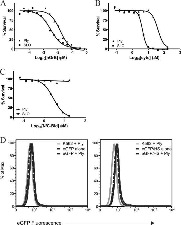
Cytotoxic lymphocytes eliminate virally infected or neoplastic cells through the action of cytotoxic proteases (granzymes). The pore-forming protein perforin is essential for delivery of granzymes into the cytoplasm of target cells; however the mechanism of this delivery is incompletely understood. Perforin contains a membrane attack complex/perforin (MACPF) domain and oligomerizes to form an aqueous pore in the plasma membrane; therefore the simplest (and best supported) model suggests that granzymes passively diffuse through the perforin pore into the cytoplasm of the target cell. Here we demonstrate that perforin preferentially delivers cationic molecules while anionic and neutral cargoes are delivered inefficiently. Furthermore, another distantly related pore-forming MACPF protein, pleurotolysin (from the oyster mushroom), also favors the delivery of cationic molecules, and efficiently delivers human granzyme B. We propose that this facilitated diffusion is due to conserved features of oligomerized MACPF proteins, which may include an anionic lumen.

Keywords: Cell Death; Cell Permeabilization; Cellular Immune Response; Granzyme; MACPF; Natural Killer (NK) Cell; Perforin; Pore; Streptolysin O; T Cell Biology.


Figures

FIGURE 1.
FIGURE 1.
Perforin preferentially delivers cationic cargo. A, K562 cells incubated with 5 μm negative (CM) or positive (DEAE) FITC-dextrans of various sizes (4–70 kDa) in the presence or absence of SLO or perforin. Representative images for delivery of positive and negative 20 kDa FITC-dextrans with SLO or perforin are shown. Images were collected at 20× magnification. B, delivery as in A of negative (black bars) or positive (white bars) FITC-dextrans with either SLO or perforin. Data are plotted as the ratio of the mean fluorescence intensity (MFI) of the dextrans in the presence of SLO or perforin (delivery), divided by the MFI of basal endocytosis. Each bar represents the mean from three independent experiments, and error bars represent standard deviation. C, native isoelectric focusing gel with a gradient of pH 3–10 loaded with 5 μg each of purified mGrB and methylated mGrB, stained with Coomassie Brilliant Blue. The pIs of the folded proteins are greater than 8.5 for mGrB (does not enter the gel) and ∼8.5 for methylated mGrB, indicated by the arrow. D, cytotoxicity of mGrB with or without methylation delivered with SLO into Jurkat cells. Data are plotted as the mean of two independent experiments for each point. D, cytotoxicity of mGrB with or without methylation delivered with perforin (data plotted as per C). E, bar graph representing the fold difference between the EC50 of unmodified mGrB and methylated mGrB. *, p < 0.05; **, p < 0.01; ***, p < 0.001 in a two tailed Student's t test.
FIGURE 2.
FIGURE 2.
Perforin facilitates delivery of cationic cytochrome c but does not deliver neutral N/C-Bid. A, 15% SDS-PAGE loaded with 5 μg each of purified cytochrome c and N/C-Bid stained with Coomassie Brilliant Blue. B, native isoelectric focusing gel with a gradient of pH 3–10 loaded with 5 μg each of purified cytochrome c and N/C-Bid and stained with Coomassie Brilliant Blue. The pIs of the folded proteins are 6.7–6.8 for N/C-Bid and higher than 8.5 for cytc. C, elution profile of cytc analyzed by size exclusion chromatography (SEC), showing that cytc elutes as one species with a single peak at 18.2 ml, indicating that it is ∼14 kDa in size as seen by SDS-PAGE in panel A. D, elution profile of N/C-Bid analyzed by SEC, showing that N/C-Bid elutes as one species with a single peak at 17.2 ml, indicating that the protein is ∼21 kDa in size, concordant with the SDS-PAGE in panel A when adding the molecular weights of the activated bid and pro-domain together. E, cytotoxicity of hGrB delivered into Jurkat cells with either SLO (squares) or perforin (circles). F, cytotoxicity of cytc delivered into P815 cells with either SLO (squares) or perforin (circles). Representative curves, n = 3 for both SLO and perforin. G, cytotoxicity of N/C-Bid when delivered into P815 cells by SLO (squares) or perforin (circles). Representative curves, n = 3 as in F. H, cytotoxicity of hGrB delivered into P815cells with perforin in the presence (squares) or absence (circles) of 32 μm N/C-Bid.
FIGURE 3.
FIGURE 3.
eGFP is not delivered by perforin and the addition of a cationic heparin binding site is not sufficient for delivery. A, 15% SDS-PAGE loaded with 5 μg of purified eGFP and eGFP/HS stained with Coomassie Brilliant Blue. B, native isoelectric focusing gel with a gradient of pH3–10 loaded with 5 μg of eGFP and eGFP/HS and stained with Coomassie Brilliant Blue. The pI of the folded eGFP was found to be 5.3, and the addition of the heparin binding site increased this to 6.3. C, representative SEC trace for eGFP. This confirmed that eGFP was monomeric and eluted at 16.5 ml, corresponding to a size of ∼30 kDa. D, heparin-Sepharose pull-down assay with eGFP versus eGFP/HS. Samples were run non-reduced and non-boiled on 12% SDS-PAGE, which was then scanned for green fluorescence. E, K562 cells were incubated with 5 μm eGFP±HS in the presence of SLO, and representative images at 40× are shown. F, representative images of delivery of eGFP±HS as per E but using perforin in place of SLO. G, delivery of eGFP±HS with SLO (n = 2) and perforin (n = 3) represented as the MFI ratio as previously described in Fig. 1B. H, cytotoxicity of hGrB when delivered into P815 cells with perforin in the presence or absence of 5 μm eGFP.
FIGURE 4.
FIGURE 4.
Positive super GFPs are delivered by perforin. A, 15% SDS-PAGE gel loaded with 5 μg each of negative 30 GFP (NEG30), standard GFP (ST), positive 15 GFP (POS15), and positive 36 GFP (POS36), stained with Coomassie Brilliant Blue. B, native isoelectric focusing gel with a gradient of pH 3–10 loaded with 5 μg of each GFP from panel (A) per well. C, SEC trace of positive 36 GFP, the single peak indicated that the protein was monomeric and pure. The positive 36 GFP (and positive 15 GFP, not shown) eluted later than expected, at ∼17.5 ml. D, 15% SDS-PAGE gel with SEC fractions under the peak from C and stained with Coomassie Brilliant Blue. This showed that the positive 36 GFP was ∼27 kDa as expected and was largely pure. E, K562 cells were incubated with 5 μm NEG30, ST, POS15, or POS36 GFP in the presence or absence of SLO (n = 2) (F) or perforin (n = 4). Data are plotted as a ratio of the MFI as previously described in Fig. 1B. *, p < 0.05; **, p < 0.01; ***, p < 0.001 in a two tailed Student's t test.
FIGURE 5.
FIGURE 5.
Pleurotolysin also preferentially delivers positively charged proteins. A, cytotoxicity of hGrB delivered into Jurkat cells with either Ply (triangles) or SLO (squares). B, cytotoxicity of cytc delivered into P815 cells with Ply (triangles) or SLO (squares). C, cytotoxicity of N/C-Bid delivered into P185 cells with Ply (triangles) or SLO (squares). D, delivery of eGFP±HS into K562 cell with Ply. FACS data represented as a histogram of eGFP fluorescence where K562 cells treated with Ply alone is in gray, K562 cells treated with eGFP±HS dotted lines, and K562 treated with eGFP±HS in the presence of Ply are represented by solid black lines.

References

    1. Peters P., Borst J., Oorschot V., Fukuda M., Krähenbühl O., Tschopp J., Slot J., Geuze H. (1991) Cytotoxic T lymphocyte granules are secretory lysosomes, containing both perforin and granzymes. J. Exp. Med. 173, 1099–1109 - PMC - PubMed
    1. Kaiserman D., Bird C. H., Sun J., Matthews A., Ung K., Whisstock J. C., Thompson P. E., Trapani J. A., Bird P. I. (2006) The major human and mouse granzymes are structurally and functionally divergent. J. Cell Biol. 175, 619–630 - PMC - PubMed
    1. Metkar S. S., Menaa C., Pardo J., Wang B., Wallich R., Freudenberg M., Kim S., Raja S. M., Shi L., Simon M. M., Froelich C. J. (2008) Human and mouse granzyme A induce a proinflammatory cytokine response. Immunity 29, 720–733 - PubMed
    1. Joeckel L. T., Wallich R., Martin P., Sanchez-Martinez D., Weber F. C., Martin S. F., Borner C., Pardo J., Froelich C., Simon M. M. (2011) Mouse granzyme K has pro-inflammatory potential. Cell Death Differ. 18, 1112–1119 - PMC - PubMed
    1. Kägi D., Ledermann B., Bürki K., Seiler P., Odermatt B., Olsen K. J., Podack E. R., Zinkernagel R. M., Hengartner H. (1994) Cytotoxicity mediated by T cells and natural killer cells is greatly impaired in perforin-deficient mice. Nature 369, 31–37 - PubMed

Publication types

LinkOut - more resources