Clinical Trial
. 2014 Apr;3(4):458-69.
doi: 10.5966/sctm.2013-0117. Epub 2014 Feb 20.

Human adult white matter progenitor cells are multipotent neuroprogenitors similar to adult hippocampal progenitors

Affiliations
Clinical Trial

Human adult white matter progenitor cells are multipotent neuroprogenitors similar to adult hippocampal progenitors

Xenia Lojewski et al. Stem Cells Transl Med. 2014 Apr.

Abstract

Adult neural progenitor cells (aNPC) are a potential autologous cell source for cell replacement in neurologic diseases or for cell-based gene therapy of neurometabolic diseases. Easy accessibility, long-term expandability, and detailed characterization of neural progenitor cell (NPC) properties are important requisites for their future translational/clinical applications. aNPC can be isolated from different regions of the adult human brain, including the accessible subcortical white matter (aNPCWM), but systematic studies comparing long-term expanded aNPCWM with aNPC from neurogenic brain regions are not available. Freshly isolated cells from subcortical white matter and hippocampus expressed oligodendrocyte progenitor cell markers such as A2B5, neuron-glial antigen 2 (NG2), and oligodendrocyte transcription factor 2 (OLIG2) in ∼20% of cells but no neural stem cell (NSC) markers such as CD133 (Prominin1), Nestin, SOX2, or PAX6. The epidermal growth factor receptor protein was expressed in 18% of aNPCWM and 7% of hippocampal aNPC (aNPCHIP), but only a small fraction of cells, 1 of 694 cells from white matter and 1 of 1,331 hippocampal cells, was able to generate neurospheres. Studies comparing subcortical aNPCWM with their hippocampal counterparts showed that both NPC types expressed mainly markers of glial origin such as NG2, A2B5, and OLIG2, and the NSC/NPC marker Nestin, but no pericyte markers. Both NPC types were able to produce neurons, astrocytes, and oligodendrocytes in amounts comparable to fetal NSC. Whole transcriptome analyses confirmed the strong similarity of aNPCWM to aNPCHIP. Our data show that aNPCWM are multipotent NPC with long-term expandability similar to NPC from hippocampus, making them a more easily accessible source for possible autologous NPC-based treatment strategies.

Keywords: Fetal neural stem cells; Hippocampus; Human adult brain; Neural progenitor cells; White matter.


Figures

Figure 1.
Figure 1.
Isolation of multipotent human aNPC from white matter and hippocampal tissue. (A): Schematic representation of experimental design and time course. (B): Representative stainings of freshly isolated cells from white matter tissue. Nuclei were counterstained with Hoechst dye. Scale bars = 100 µm. (C): Quantitative analysis of marker protein expression in freshly isolated cells from white matter or hippocampal tissue. No cells with marker expression were detected for CD133 (Prominin1), SOX2, PAX6, GALC, O4, GFAP, S100β, glutamate aspartate transporter, polysialylated neural cell adhesion protein Vimentin, and fibronectin. ∗, p < .05 when compared with white matter tissue (two-sided t test). (D): Quantitative transcription profile of the tissue samples. qRT-PCR analyses were performed for marker genes of neural stem cells (SOX2, MSI1, NESTIN, MKI67), glial lineage (CSPG4, OLIG2, PDGFRα, GALC, GFAP), and neuronal lineage-restricted progenitors (SOX1, PAX6, TUBB3). Results are mean values ± SEM from at least three independent experiments. ∗, p < .05 when compared with white matter tissue (two-sided t test). Limiting dilution assay for primary neurospheres derived from white matter (E) and hippocampus (F). Data are shown as mean ± SEM (n = 3–5), and dotted lines represent the 95% confidence interval (95% CI). The intercept of log (37% negative wells) gave the neurosphere-forming frequency. Insets show representative photographs of respective primary neurosphere. Note the statistical significant difference in neurosphere-forming capacity between the two tissues. Scale bars = 100 µm. Abbreviations: CI, confidence interval; EGFR, epidermal growth factor receptor; GALC, galactocerebroside; GFAP, glial fibrillary acidic protein; HMBS, hydroxymethylbilane synthase; NG2, neuron-glial antigen 2 (chondroitin sulfate proteoglycan 4); OLIG2, oligodendrocyte transcription factor 2; PDGFR, platelet-derived growth factor receptor; qRT-PCR, quantitative reverse transcription-polymerase chain reaction.
Figure 2.
Figure 2.
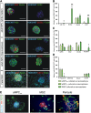
Expansion of multipotent human adult neural progenitor cells (aNPC) from white matter and hippocampal tissue. Human aNPC from white matter and hippocampus were cultured in suspension (neurosphere) culture for up to 6 weeks and stained for various marker proteins. aNPC were compared with human fetal neural stem cell (NSC) as a multipotent NSC control. (A): Representative selection of stainings for typical marker proteins in aNPCWM, aNPCHIP, and fNSC. Nuclei were counterstained with Hoechst dye. Scale bars = 100 µm. (B–D): Quantitative analysis of marker proteins. Cells were stained against NSC and proliferation markers (CD133, SOX2, Nestin, KI67), markers for oligodendroglial progenitor cells (A2B5, NG2, OLIG2, PDGFRα), and differentiated cells (β-Tubulin III, MAP2ab, GALC, GFAP). One-way analysis of variance revealed statistical significant differences for CD133 (F value: 12.00; p = .008), SOX2 (F value: 59.31; p < .001), KI67 (F value: 17.16; p < .001), NG2 (F value: 15.85; p = .002), OLIG2 (F value: 4.24; p = .043), PDGFRα (F value: 6.46; p = .032), and β-Tubulin III (F value: 7.33; p = .011). Results are mean values ± SEM from at least three independent experiments. ∗, p < .05; ∗∗, p < .01; ∗∗∗, p < .001 when compared with aNPCWM (post hoc Bonferroni adjusted t test). #, p < .05; ##, p < .01; ###, p < .001 when compared with aNPCHIP (post hoc Bonferroni adjusted t test). (E): aNPC do not express the pericyte marker SMA. Depicted are representative images of aNPC, MSC, and adult human brain-derived pericytes. Magnification, ×200. Abbreviations: aNPCHIP, hippocampal adult neural progenitor cell; aNPCWM, white matter adult neural progenitor cell; fNSC, fetal neural stem cell; GALC, galactocerebroside; GFAP, glial fibrillary acidic protein; MAP2, mitogen-activated protein 2; MSC, mesenchymal stem cell; NG2, neuron-glial antigen 2; OLIG2, oligodendrocyte transcription factor 2; PDGFR, platelet-derived growth factor receptor; TubIII, β-Tubulin III.
Figure 3.
Figure 3.
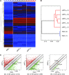
Characterization of multipotent adult neural progenitor cells (aNPC) from white matter and hippocampal tissue. (A): Heat map from microarray data demonstrating global gene expression pattern of white matter and hippocampal aNPC in comparison with fNSC. The color bar at the top indicates gene expression in log2 scale. Red and blue colors represent higher and lower gene expression levels, respectively. (B): Hierarchical clustering of the cell lines based on the gene expression profiles in (A). (C): Pairwise scatter plot analysis of the global gene expression profiles of aNPC and fNSC. Black lines indicate boundaries of twofold difference in gene expression levels. The positions of some markers are shown as orange dots. Gene expression levels are depicted in log2 scale. The number of differentially expressed genes is indicated under each scatter plot. Abbreviations: aNPCHIP, hippocampal adult neural progenitor cell; aNPCWM, white matter adult neural progenitor cell; diff., differentially expressed; fNSC, fetal neural stem cell; OLIG2, oligodendrocyte transcription factor 2.
Figure 4.
Figure 4.
Quantitative reverse transcription-polymerase chain reaction characterization of multipotent human adult neural progenitor cells (aNPC) from white and hippocampal tissue. Gene expression analysis of marker genes for neural stem/progenitor cells (A), glial lineage-restricted cells (B), and neuronal restricted progenitor cells (C) in aNPC and fNSC during expansion using quantitative reverse transcription-polymerase chain reaction. One-way analysis of variance revealed statistical significant differences for SOX2 (F value: 6.6; p = .015), MSI1 (F value: 11.0; p = .004), MKI67 (F value: 20.4; p < .001), PDGFRα (F value: 35.4; p < .001), PAX6 (F value: 6.6; p = .025), and TUBIII (F value: 5.3; p = .040), but not for CD133 (F value: 0.2; p = .805), NESTIN (F value: 3.3; p = .086), CSPG4 (F value: 1.6; p = .248), OLIG2 (F value: 0.9; p = .437), GALC (F value: 1.1; p = .377), GFAP (F value: 3.4; p = .091), and SOX1 (F value: 152; p value <.0001). Results are mean values ± SEM from at least three independent experiments. ∗, p < .05; ∗∗, p < .01; ∗∗∗, p < .001 when compared with aNPCWM (post hoc Bonferroni adjusted t test). #, p < .05; ##, p < .01; ###, p < .001 when compared with aNPCHIP (post hoc Bonferroni adjusted t test). Abbreviations: aNPCHIP, hippocampal adult neural progenitor cell; aNPCWM, white matter adult neural progenitor cell; fNSC, fetal neural stem cell; GALC, galactocerebroside; GFAP, glial fibrillary acidic protein; HMBS, hydroxymethylbilane synthase; MSI1, Musashi1; OLIG2, oligodendrocyte transcription factor 2; PDGFR, platelet-derived growth factor receptor.
Figure 5.
Figure 5.
Differentiation of multipotent human adult neural progenitor cell from white matter and hippocampal tissue. Human adult neural progenitor cells (NPC) were cultured in suspension (neurosphere) culture for 6 weeks, differentiated for 14 days, and stained for various marker proteins. Adult NPC were compared with human fetal neural stem cells (NSC) as a multipotent NSC control. (A): Representative selection of stainings. Cells were stained against markers for neurons (β-Tubulin III, MAP2ab), oligodendrocytes (GALC), and astrocytes (GFAP). Nuclei were counterstained with Hoechst dye. Scale bars = 100 µm. (B): Quantitative analysis of marker proteins in 14-day-old cultures, as shown in (A). One-way analysis of variance revealed statistical significant differences only for MAP2ab (F value: 53.1; p < .001) and GALC (F value: 4.9; p = .047), but not for β-Tubulin III (F value: 2.0; p = .185) and GFAP (F value: 2.7; p = .119). Results are mean values ± SEM from at least three independent experiments. ∗, p < .05; ∗∗∗, p < .001 when compared with aNPCWM (post hoc Bonferroni adjusted t test). #, p < .05; ###, p < .001 when compared with aNPCHIP (post hoc Bonferroni adjusted t test). (C–E): Electrophysiological data of human aNPCWM after differentiation for 4 weeks in vitro. (C): Whole-cell voltage clamp recordings of a sodium inward current (negative current) induced by stepwise depolarization with increasing amplitudes from a holding potential of −70 mV. (D): Current voltage plots of NPC showing the mean amplitudes of sodium inward and potassium outward currents. (E): Representative action potential recording of a differentiated NPC in response to a depolarizing current pulse; the holding potential was −65 mV in current-clamp mode. Abbreviations: aNPCHIP, hippocampal adult neural progenitor cell; aNPCWM, white matter adult neural progenitor cell; fNSC, fetal neural stem cell; GALC, galactocerebroside; GFAP, glial fibrillary acidic protein; MAP2, mitogen-activated protein 2; TubIII, β-Tubulin III.

Similar articles

Cited by

References

    1. Hermann A, Maisel M, Liebau S, et al. Mesodermal cell types induce neurogenesis from adult human hippocampal progenitor cells. J Neurochem. 2006;98:629–640. - PubMed
    1. Coras R, Siebzehnrubl FA, Pauli E, et al. Low proliferation and differentiation capacities of adult hippocampal stem cells correlate with memory dysfunction in humans. Brain. 2010;133:3359–3372. - PubMed
    1. Kukekov VG, Laywell ED, Suslov O, et al. Multipotent stem/progenitor cells with similar properties arise from two neurogenic regions of adult human brain. Exp Neurol. 1999;156:333–344. - PubMed
    1. Nunes MC, Roy NS, Keyoung HM, et al. Identification and isolation of multipotential neural progenitor cells from the subcortical white matter of the adult human brain. Nat Med. 2003;9:439–447. - PubMed
    1. Scolding NJ, Rayner PJ, Compston DA. Identification of A2B5-positive putative oligodendrocyte progenitor cells and A2B5-positive astrocytes in adult human white matter. Neuroscience. 1999;89:1–4. - PubMed

Publication types

Substances