. 2013 Jan 1;40(12):1748-1758.
doi: 10.1080/02678292.2013.846422.

Liquid crystal assemblies in biologically inspired systems

Affiliations

Liquid crystal assemblies in biologically inspired systems

Cyrus R Safinya et al. Liq Cryst. .

Abstract

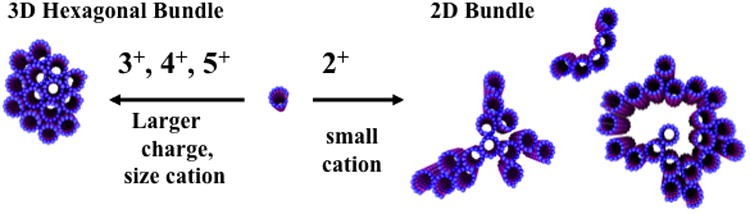
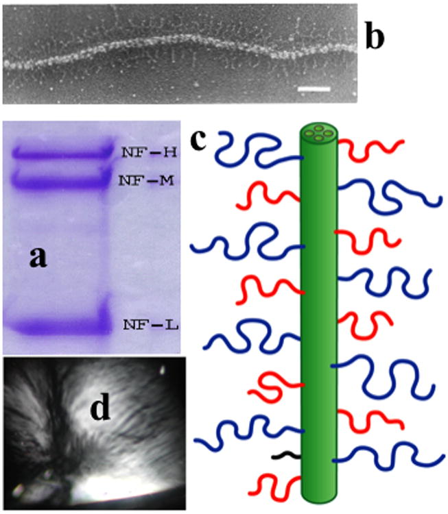
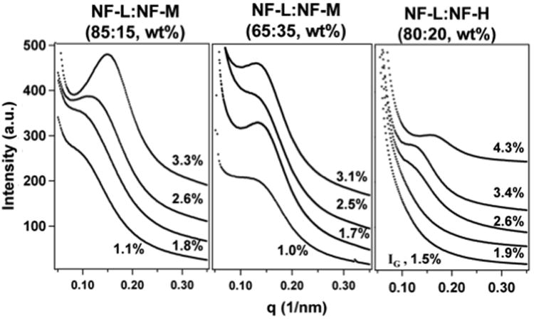
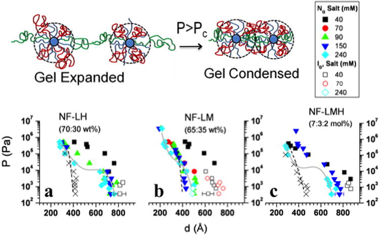
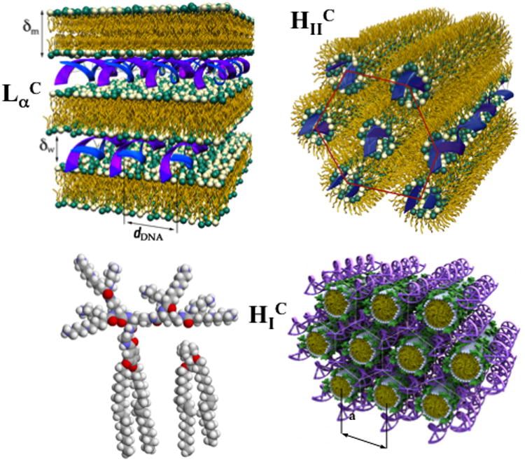
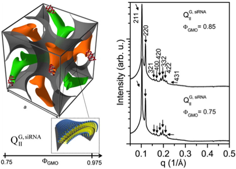
In this paper, which is part of a collection in honor of Noel Clark's remarkable career on liquid crystal and soft matter research, we present examples of biologically inspired systems, which form liquid crystal (LC) phases with their LC nature impacting biological function in cells or being important in biomedical applications. One area focuses on understanding network and bundle formation of cytoskeletal polyampholytes (filamentous-actin, microtubules, and neurofilaments). Here, we describe studies on neurofilaments (NFs), the intermediate filaments of neurons, which form open network nematic liquid crystal hydrogels in axons. Synchrotron small-angle-x-ray scattering studies of NF-protein dilution experiments and NF hydrogels subjected to osmotic stress show that neurofilament networks are stabilized by competing long-range repulsion and attractions mediated by the neurofilament's polyampholytic sidearms. The attractions are present both at very large interfilament spacings, in the weak sidearm-interpenetrating regime, and at smaller interfilament spacings, in the strong sidearm-interpenetrating regime. A second series of experiments will describe the structure and properties of cationic liposomes (CLs) complexed with nucleic acids (NAs). CL-NA complexes form liquid crystalline phases, which interact in a structure-dependent manner with cellular membranes enabling the design of complexes for efficient delivery of nucleic acid (DNA, RNA) in therapeutic applications.

Keywords: DNA; Neurofilaments; RNA; gyroid cubic phases; hexagonal liquid crystals; lamellar liquid crystals; lipids; nematic hydrogels; small-angle-x-ray-scattering (SAXS).


Figures

Figure 1
Figure 1
Schematic of higher-order-assembly of microtubules in the presence of multivalent counterions. (Left) Trivalent (spermidine [H3N+-(CH2)3-+NH2-(CH2)4-+NH3] and lysine3), tetravalent (spermine [H3N+-(CH2)3-+NH2-(CH2)4-+NH2-(CH2)3-+NH3] and lysine4), and pentavalent (lysine5) cations lead to the formation of 3D bundles with hexagonal in-plane symmetry. (Right) Divalent cations [(Ba2+, Ca2+, Sr2+)] lead to the sheet-like 2D bundles with linear, branched, and loop morphologies. Reprinted with permission from [17]. Copyright 2004, National Academy of Sciences, U.S.A.
Figure 2
Figure 2
Quick-frozen, deep-etched electron micrograph of a mouse nerve axon showing a microtubule (MT, red arrow) with an attached vesicle moving along the MT track via molecular motors. The MT is seen to be immersed in a network of Neurofilaments (NFs, red stars). The NF-networks in vertebrate axons form a nematic liquid crystal hydrogel. The scale in this micrograph can be seen by noting that the diameter of a MT is 25 nm. Reprinted from [7], with permission from Elsevier.
Figure 3
Figure 3
(a) Gel electrophoresis (10% polyacrylamide in sodium dodecyl sulfate) of purified neurofilaments (NFs) showing the three subunits (NF-L, MW = 60K; NF-M, MW = 100K; NF-H, MW = 115K). NFs purified from bovine spinal cord according to the procedures described in [23, 25, 27]. (b) Electron micrograph of a mature neurofilament (glycerol sprayed/low-angle rotary metal shadowed on freshly cleaved mica). The sidearms are clearly visible. The bar is 100 nm. (c) Schematic of a NF showing the sidearms of NF-H (blue), NF-M (red), and NF-L (black) subunits containing 613, 514, and 158, amino acid residues respectively. (d) A reconstituted NF mixture forming a strongly birefringent hydrogel viewed between crossed polarizers shows the presence of nematic-like liquid crystalline texture. Parts a and d reprinted with permission from [22]; part b reprinted with permission from [6].
Figure 4
Figure 4
Synchrotron SAXS data of NF-LM and NF-LH neurofilaments assembled from NF-L and NF-M (Left and Middle) and NF-L and NF-H (Right) subunits in 86 mM monovalent salt and pH ≈ 6.8. Each figure shows data along dilution lines (with decreasing total protein concentrations) for NF-L/NF-M = 85/15 (wt/wt), NF-L/NF-M = 65/35 (wt/wt) and NF-L/NF-H = 80/20 (wt/wt). Reprinted from [23], with permission from the Biophysical Society.
Figure 5
Figure 5
(Top) End view of NF-LH, NF-LMH hydrogels undergoing a transition for P > Pc from an expended-network to a condensed-network gel state with interpenetrating sidearm polyampholyte chains. (Bottom) Pressure-interfilament distance curves for NF hydrogels obtained using the SAXS-osmotic pressure technique. At ionic strengths 150 mM and 240 mM NF-LH (a) and NF-LMH (c) gels exhibit an expanded-network to condensed-network transition for P > Pc ≈ 104 Pa, whereas NF-LM gels (b) are in a compressed state for P < Pc and transition to the condensed state for P > Pc. (NG = nematic gel; IG = isotropic gel). At low ionic strengths (40 mM and 70 mM) NF-LM gels are expanded isotropic gels at P < Pc and transition to the condensed-network state at P > Pc. Reprinted in part with permission from [25].
Figure 6
Figure 6
The structure of three distinct cationic liposome (CL)-DNA complexes determined by synchrotron small-angle x-ray scattering. (Top left) The structure of the lamellar LαC phase of CL-DNA complexes with DNA sandwiched between lipid bilayers. The interlayer spacing is d = δw + δm. (Top right) The structure of the inverted hexagonal HIIC phase of CL-DNA complexes comprised of DNA coated with a lipid monolayer arranged on a hexagonal lattice. (Bottom left) Molecular models of hexadecavalent lipid MVLBG2 (16+) and monovalent lipid 1,2-dioleoyl-3-trimethylammonium-propane (DOTAP, 1+). (Bottom right) Schematic of the HIC phase of CL-DNA complexes where the lipids consist of a mixture of MVLBG2 and 1,2-dioleoyl-sn-glycero-3-phosphocholine (DOPC). The large headgroup of MVLBG2 forces the lipid to assemble into cylindrical lipid micelles that are arranged on a hexagonal lattice. The DNA rods are arranged on a honeycomb lattice in the interstices of the lipid micelle arrangement. The interstitial space is filled by the headgroups, water and counterions. Interestingly, the DNA forms a three-dimensionally contiguous substructure in the HIC phase, as opposed to isolated DNA rods and sheets of parallelly arranged strands in the HIIC and LαC phases, respectively. Bottom images reprinted with permission from [36]. Copyright 2006 American Chemical Society. Top images reprinted with permission from [35].
Figure 7
Figure 7
(Left) The double gyroid lipid cubic phase incorporating functional short-interfering RNA (siRNA) within its two (green and orange) water channels. This phase, labeled QIIG,siRNA, is obtained for DOTAP/GMO-siRNA complexes with 1-monooleoyl-glycerol (GMO) molar fractions (ΦGMO) of 0.75 ≤ ΦGMO ≤ 0.975. A lipid bilayer surface separates the two intertwined but independent water channels. For clarity, the bilayer (which has a negative Gaussian curvature C1C2 < 0) is represented by a surface (grey) corresponding to a thin layer in the center of the membrane as indicated in the enlarged inset. (Right) Synchrotron small-angle X-ray scattering data for DOTAP/GMO in 30 wt% water containing siRNA molecules at ΦGMO = 0.75 (below) and at ΦGMO = 0.85 (above). The large number of reflections for the QIIG,siRNA phase result from a body centered gyroid cubic structure with space group Ia3d. Reprinted with permission from [37]. Copyright 2010 American Chemical Society.

Similar articles

Cited by

References

    1. Kandel ER, Schwartz JH, Jessell TM. Principles of Neural Science. 4th. McGraw Hill; New York: 2000.
    1. Peters A, Palay SL, Def H. The Fine Structure of the Nervous System. 3rd. Webster; New York: 1991.
    1. Burgoyne RD, editor. The Neuronal cytoskeleton. Wiley & Sons; New York: 1991.
    1. Hirokawa N. In: The Neuronal Cytoskeleton. Burgoyne RD, editor. Wiley; New York: 1991. pp. 5–74.
    1. Hirokawa N. Cross-linker system between neurofilaments, microtubules and membranous organelles in frog axons revealed by the quick-freeze, deep-etching method. J Cell Biol. 1982;94:129–142. - PMC - PubMed