A novel radiation-induced p53 mutation is not implicated in radiation resistance via a dominant-negative effect
- PMID: 24558369
- PMCID: PMC3928108
- DOI: 10.1371/journal.pone.0087492
A novel radiation-induced p53 mutation is not implicated in radiation resistance via a dominant-negative effect
Abstract
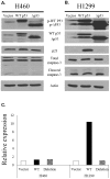
Understanding the mutations that confer radiation resistance is crucial to developing mechanisms to subvert this resistance. Here we describe the creation of a radiation resistant cell line and characterization of a novel p53 mutation. Treatment with 20 Gy radiation was used to induce mutations in the H460 lung cancer cell line; radiation resistance was confirmed by clonogenic assay. Limited sequencing was performed on the resistant cells created and compared to the parent cell line, leading to the identification of a novel mutation (del) at the end of the DNA binding domain of p53. Levels of p53, phospho-p53, p21, total caspase 3 and cleaved caspase 3 in radiation resistant cells and the radiation susceptible (parent) line were compared, all of which were found to be similar. These patterns held true after analysis of p53 overexpression in H460 cells; however, H1299 cells transfected with mutant p53 did not express p21, whereas those given WT p53 produced a significant amount, as expected. A luciferase assay demonstrated the inability of mutant p53 to bind its consensus elements. An MTS assay using H460 and H1299 cells transfected with WT or mutant p53 showed that the novel mutation did not improve cell survival. In summary, functional characterization of a radiation-induced p53 mutation in the H460 lung cancer cell line does not implicate it in the development of radiation resistance.
Conflict of interest statement
Figures


Similar articles
-
WTp53 induction does not override MTp53 chemoresistance and radioresistance due to gain-of-function in lung cancer cells.Mol Cancer Ther. 2008 Apr;7(4):980-92. doi: 10.1158/1535-7163.MCT-07-0471. Mol Cancer Ther. 2008. PMID: 18413811
-
Radiosensitization of Non-Small Cell Lung Cancer Cells by Inhibition of TGF-β1 Signaling With SB431542 Is Dependent on p53 Status.Oncol Res. 2016;24(1):1-7. doi: 10.3727/096504016X14570992647087. Oncol Res. 2016. PMID: 27178816 Free PMC article.
-
Micheliolide Enhances Radiosensitivities of p53-Deficient Non-Small-Cell Lung Cancer via Promoting HIF-1α Degradation.Int J Mol Sci. 2020 May 11;21(9):3392. doi: 10.3390/ijms21093392. Int J Mol Sci. 2020. PMID: 32403326 Free PMC article.
-
Clinical implication of p53 mutation in lung cancer.Mol Biotechnol. 2003 Jun;24(2):141-56. doi: 10.1385/MB:24:2:141. Mol Biotechnol. 2003. PMID: 12746555 Review.
-
P53-mediated radioresistance does not correlate with metastatic potential in tumorigenic rat embryo cell lines following oncogene transfection.Int J Radiat Oncol Biol Phys. 1996 Jan 15;34(2):341-55. doi: 10.1016/0360-3016(95)02023-3. Int J Radiat Oncol Biol Phys. 1996. PMID: 8567335 Review.
Cited by
-
p53 Modulates Radiosensitivity in Head and Neck Cancers-From Classic to Future Horizons.Diagnostics (Basel). 2022 Dec 5;12(12):3052. doi: 10.3390/diagnostics12123052. Diagnostics (Basel). 2022. PMID: 36553058 Free PMC article. Review.
References
-
- Hemminki K, Lenner P, Sundquist J, Bermejo JL (2008) Risk of subsequent solid tumors after non-hodgkin's lymphoma: Effect of diagnostic age and time since diagnosis. J Clin Oncol 26 (11) 1850–18577. - PubMed
-
- Ettinger DS, Akerley W, Borghaei H, Chang AC, Cheney RT, et al. (2013) Non-small cell lung cancer, version 2. J Natl Compr Canc Netw 11 (6) 645. - PubMed
-
- Huang C (ed) (2012) Protein phosphorylation in human Health.
-
- Haupt S, Berger M, Goldberg Z, Haupt Y (2003) Apoptosis - the p53 network. J Cell Sci 16: 4077–4085. - PubMed
-
- Schuler M, Green DR (2001) Mechanisms of p53-dependent apoptosis. Biochem Soc Trans 29: 684–688. - PubMed
Publication types
MeSH terms
Substances
Grants and funding
LinkOut - more resources
Full Text Sources
Other Literature Sources
Medical
Research Materials
Miscellaneous

