Comparative Study
. 2014 Feb 18;9(2):e88840.
doi: 10.1371/journal.pone.0088840. eCollection 2014.

To nick or not to nick: comparison of I-SceI single- and double-strand break-induced recombination in yeast and human cells

Affiliations
Comparative Study

To nick or not to nick: comparison of I-SceI single- and double-strand break-induced recombination in yeast and human cells

Samantha S Katz et al. PLoS One. .

Abstract

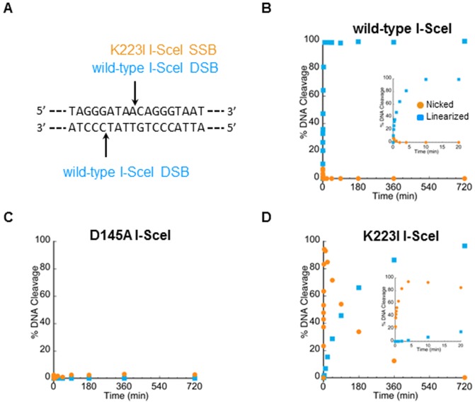
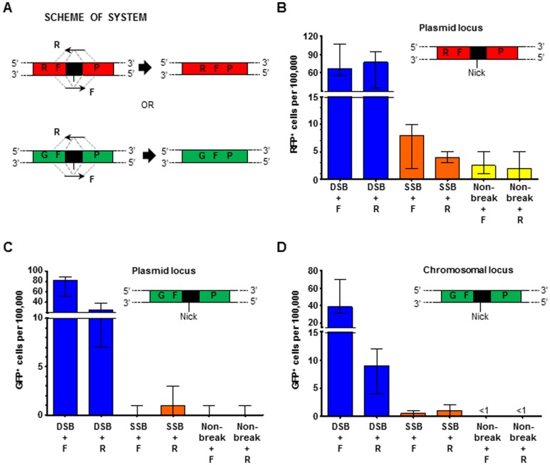
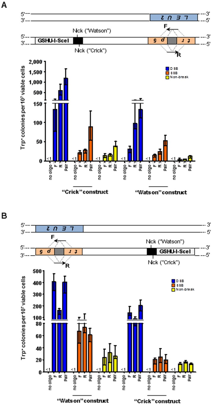
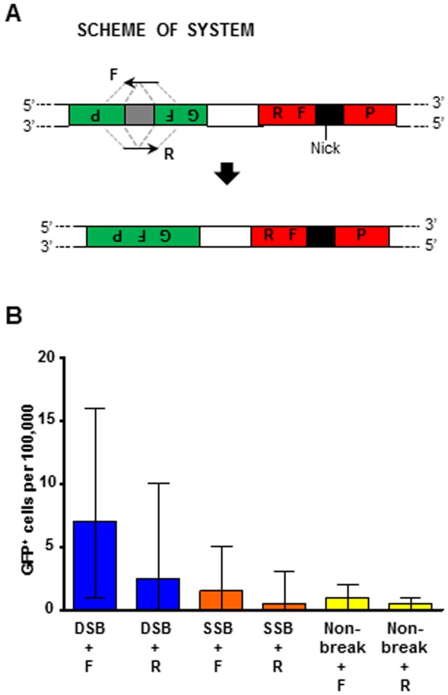
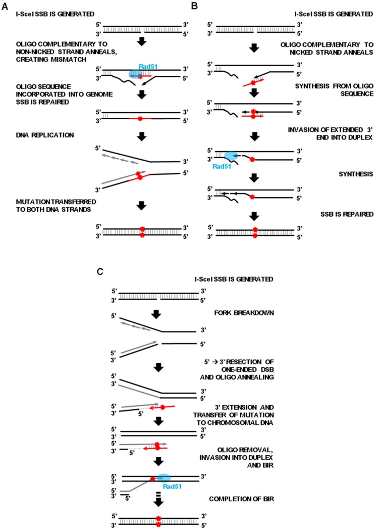
Genetic modification of a chromosomal locus to replace an existing dysfunctional allele with a corrected sequence can be accomplished through targeted gene correction using the cell's homologous recombination (HR) machinery. Gene targeting is stimulated by generation of a DNA double-strand break (DSB) at or near the site of correction, but repair of the break via non-homologous end-joining without using the homologous template can lead to deleterious genomic changes such as in/del mutations, or chromosomal rearrangements. By contrast, generation of a DNA single-strand break (SSB), or nick, can stimulate gene correction without the problems of DSB repair because the uncut DNA strand acts as a template to permit healing without alteration of genetic material. Here, we examine the ability of a nicking variant of the I-SceI endonuclease (K223I I-SceI) to stimulate gene targeting in yeast Saccharomyces cerevisiae and in human embryonic kidney (HEK-293) cells. K223I I-SceI is proficient in both yeast and human cells and promotes gene correction up to 12-fold. We show that K223I I-SceI-driven recombination follows a different mechanism than wild-type I-SceI-driven recombination, thus indicating that the initial DNA break that stimulates recombination is not a low-level DSB but a nick. We also demonstrate that K223I I-SceI efficiently elevates gene targeting at loci distant from the break site in yeast cells. These findings establish the capability of the I-SceI nickase to enhance recombination in yeast and human cells, strengthening the notion that nicking enzymes could be effective tools in gene correction strategies for applications in molecular biology, biotechnology, and gene therapy.


Conflict of interest statement

Competing Interests: The authors have declared that no competing interests exist.

Figures

Figure 1
Figure 1. DNA cleavage activities of the wild-type, D145A, and K223I I-SceI proteins.
(A) Scheme of the 18-bp I-SceI recognition sequence showing the cleavage positions of wild-type I-SceI and K223I I-SceI. Supercoiled pBS-I-SceI (E/H) plasmid DNA was incubated with (B) wild-type I-SceI, (C) D145A I-SceI mutant, or (D) K223I I-SceI mutant for various lengths of time and the amounts of the nicked open circle (orange circles) and linear (blue squares) reaction product DNAs were plotted as a function of time. Data points represent the average values of two experiments. Insets show the same data immediately following initiation of the reactions.
Figure 2
Figure 2. An I-SceI K223I break stimulates HR between direct repeats in yeast.
(A) Scheme showing disrupted yeast lys2 chromosomal locus containing the I-SceI recognition sequence (black box) within 90-bp direct repeats (small arrows). The position of the SSB is indicated (“Nick”). DNA strands are identified as “Crick” and “Watson” according to the Saccharomyces cerevisiae Genome Database (SGD). (B) Frequency of Lys+ recombinants following expression of wild-type I-SceI (dark blue bars labeled “DSB”), K223I I-SceI (orange bars labeled “SSB”), or D145A I-SceI (yellow bars labeled “Non-break”) in RAD51 wild-type (left) or rad51-null mutant (right) strains are presented as the median with range (n≥11). For the specific numerical values see Table S3A. Wild-type I-SceI strains used: SAS-74 and SAS-75 (RAD51), and SAS-174 and SAS-175 (rad51Δ). K223I I-SceI strains used: SAS-77 and SAS-149 (RAD51), and SAS-176 and SAS-177 (rad51Δ). D145A I-SceI strains used: SAS-142 and SAS-143 (RAD51), and SAS-178 and SAS-179 (rad51Δ).
Figure 3
Figure 3. A K223I I-SceI break stimulates gene correction by oligonucleotides in yeast.
(A) Scheme showing disrupted yeast trp5 chromosomal locus containing the I-SceI recognition sequence (black box). The position of the SSB is indicated (“Nick”) for the “Crick” and “Watson” constructs. Dashed gray lines indicate the complementarity between the F oligonucleotide and the antisense strand of the targeted gene, and between the R oligonucleotide and the sense strand of the targeted gene. (B–D) Frequencies of Trp+ transformants following expression of wild-type I-SceI (dark blue bars labeled “DSB”), K223I (orange bars labeled “SSB”), or D145A (yellow bars labeled “Non-break”) using either of the single or the pair of oligonucleotides to repair the break. All data are presented as the median with range (n≥5). For the specific numerical values see Table S3B-D. (B) Gene correction frequencies by oligonucleotides when an SSB is generated on the “Crick” (left) or “Watson” (right) chromosomal strand. (C) Frequency of transformants in RAD51 (left) or rad51 null mutant (right) strains when the SSB is generated on the “Crick” strand. (D) Frequency of transformants following expression of wild-type I-SceI in RAD51 (left) or rad51 null mutant (right) strains with final galactose concentrations of 2% (dark blue bars labeled “DSB (2%)”) or 0.02% (light blue bars labeled “DSB (0.02%)”). Wild-type I-SceI strains used: SAS-227 and SAS-228 (“Crick” and RAD51), SAS-281 and SAS-282 (“Watson”), and SAS-235 and SAS-236 (rad51Δ). K223I I-SceI strains used: SAS-229 and SAS-230 (“Crick” and RAD51), SAS-283 and SAS-284 (“Watson”), and SAS-237 and SAS-238 (rad51Δ). D145A I-SceI strains used: SAS-231 and SAS-232 (“Crick” and RAD51), SAS-285 and SAS-286 (“Watson”), and SAS-239 and SAS-240 (rad51Δ).
Figure 4
Figure 4. A nick occurring in asynchronous or G1 arrested cells stimulates gene correction by oligonucleotides equally efficiently.
Shown are frequency of Trp+ transformants by oligonucleotides when cells were asynchronous (left) or arrested in G1 (right) at the time of wild-type-I-SceI or K223I I-SceI breakage prior to oligonucleotide transformation and when the SSB is generated on the “Crick” strand. All data are presented as the median with range (n≥5). For the specific numerical values see Table S3E. Wild-type I-SceI strains used: SAS-227 and SAS-228. K223I I-SceI strains used: SAS-229 and SAS-230. D145A I-SceI strains used: SAS-231 and SAS-232.
Figure 5
Figure 5. K223I I-SceI SSB can stimulate recombination in human cells.
(A) Scheme showing disrupted RFP plasmid locus and disrupted GFP plasmid or chromosomal locus containing the I-SceI recognition sequence (black box). The position of the SSB is indicated by a black line. Dashed gray lines indicate the complementarity between the F oligonucleotide and the antisense strand of the targeted gene, and between the R oligonucleotide and the sense strand of the targeted gene. (B–D) Frequency of fluorescent cells following expression of wild-type I-SceI (dark blue bars labeled “DSB”), K223I I-SceI (orange bars labeled “SSB”), or D145A I-SceI (yellow bars labeled “Non-break”) using either of the single oligonucleotides to repair the break. All data are presented as the median with range. For the specific numerical values see Table S3F-H. (B) Recombination at the RFP target plasmid locus (n = 6). (C) Recombination at the GFP target plasmid locus (n = 9). (D) Recombination at the GFP target chromosomal locus (n≥8).
Figure 6
Figure 6. Gene targeting distant from the I-SceI DSB or SSB in yeast cells.
Depicted above each graph in (A) and (B) are schemes of the two copies of chromosome VII of diploid yeast cells, one in which TRP5 has been replaced by LEU2, and another in which TRP5 has been disrupted by a 31-bp insertion (gray box), which does not contain an I-SceI cut site, and which also contains the GSHU-I-SceI cassette with the I-SceI site (black box) inserted 10 kb upstream or downstream of the disrupted trp5. Dashed gray lines indicate the complementarity between the F oligonucleotide and the antisense strand of the targeted gene, and between the R oligonucleotide and the sense strand of the targeted gene. The position of the SSB is indicated (“Nick”) for the “Watson” and “Crick” constructs. Frequencies of Trp+ transformants following expression of wild-type I-SceI (dark blue bars labeled “DSB”), K223I I-SceI (orange bars labeled “SSB”), or D145A I-SceI (yellow bars labeled “Non-break”) using either of the single oligonucleotides or the pair of oligonucleotides to repair the break. All data are presented as the median with range (n≥5). For the specific numerical values see Table S3I,J. (A) Frequencies of transformants when an SSB is generated on the “Crick” (left) or “Watson” (right) chromosomal strand 10 kb upstream from the trp5 locus. Wild-type I-SceI strains used: SAS-150 and SAS-151 (“Crick”), and SAS-215 and SAS-217 (“Watson”). K223I I-SceI strains used: SAS-162 and SAS-163 (“Crick”), and SAS-207 and SAS-209 (“Watson”). D145A I-SceI strains used: SAS-166 and SAS-167 (“Crick”), and SAS-211 and SAS-213 (“Watson”). (B) Frequencies of transformants when an SSB is generated on the top (“Watson”, left) or bottom (“Crick”, right) chromosomal strand 10 kb downstream from the trp5 locus. Wild-type I-SceI strains used: SAS-152 and SAS-153 (“Watson”), and SAS-272 and SAS-274 (“Crick”). K223I I-SceI strains used: SAS-154 and SAS-156 (“Watson”), and SAS-219 and SAS-221 (“Crick”). D145A I-SceI strains used: SAS-158 and SAS-160 (“Watson”), and SAS-251 and SAS-253 (“Crick”).
Figure 7
Figure 7. Gene targeting distant from the I-SceI DSB or SSB in human cells.
(A) Scheme showing the disrupted GFP plasmid locus 2.3 kb distant from the I-SceI recognition sequence (black box). The position of the SSB is indicated (“Nick”). Dashed gray lines indicate the complementarity between the F oligonucleotide and the antisense strand of the targeted gene, and between the R oligonucleotide and the sense strand of the targeted gene. (B) Frequencies of GFP+ cells following expression of wild-type I-SceI (dark blue bars labeled “DSB”), K223I I-SceI (orange bars labeled “SSB”), or D145A I-SceI (yellow bars labeled “Non-break”) using either of the single oligonucleotides to correct the GFP gene distant from the I-SceI break. All data are presented as the median with range (n = 6). For the specific numerical values see Table S3K.
Figure 8
Figure 8. Models for I-SceI K223I SSB-driven HR using single-stranded oligonucleotides.
(A) Gene correction using an oligonucleotide complementary to the intact strand. After the SSB is generated, an oligonucleotide (red arrow) carrying the desired nucleotide change (red star) anneals to the complementary strand by invading the nicked duplex with the help of Rad51. The oligonucleotide is incorporated in the duplex and its genetic modification is transferred to the other strand in the subsequent round of DNA replication. (B) Gene correction using an oligonucleotide complementary to the nicked strand. After the SSB is generated, an oligonucleotide sequence (red arrow) with the desired mutation (red star) can serve as template to extend the unwound 3′ broken end. This extended 3′ end may invade the duplex via Rad51 function. The 3′ end is then extended further. The nick is repaired and the mutation fixed by mismatch repair or in the next round of replication. (C) Gene correction following collapse of a replication fork at the nick. After the SSB is generated, it persists until encountered by the replication fork. Following fork breakdown, a one-ended DSB forms. Resection of the 5′ end (indicated by a light gray dashed line) produces a single-strand 3′ end that anneals with the complementary sequence of an oligonucleotide (red arrow) containing a desired mutation (red star) and uses the oligonucleotide as a template for extension. After removal of the oligonucleotide by unwinding or resection, the 3′ end (dark gray) invades the intact duplex via Rad51 and follows the steps (dotted black arrow) of BIR to complete repair. The mutation carried on the 3′end is passed to the other strand of the chromosome by mismatch repair or in the next round of replication.

References

    1. Capecchi MR (1989) Altering the genome by homologous recombination. Science 244: 1288–1292. - PubMed
    1. Hinnen A, Hicks JB, Fink GR (1978) Transformation of yeast. Proc Natl Acad Sci U S A 75: 1929–1933. - PMC - PubMed
    1. Orr-Weaver TL, Szostak JW, Rothstein RJ (1981) Yeast transformation: a model system for the study of recombination. Proc Natl Acad Sci U S A 78: 6354–6358. - PMC - PubMed
    1. Doetschman T, Gregg RG, Maeda N, Hooper ML, Melton DW, et al. (1987) Targetted correction of a mutant HPRT gene in mouse embryonic stem cells. Nature 330: 576–578. - PubMed
    1. Robertson E, Bradley A, Kuehn M, Evans M (1986) Germ-line transmission of genes introduced into cultured pluripotential cells by retroviral vector. Nature 323: 445–448. - PubMed

Publication types