. 2014 Feb 18;9(2):e88910.
doi: 10.1371/journal.pone.0088910. eCollection 2014.

Long-term activation upon brief exposure to xanomleline is unique to M1 and M4 subtypes of muscarinic acetylcholine receptors

Affiliations

Long-term activation upon brief exposure to xanomleline is unique to M1 and M4 subtypes of muscarinic acetylcholine receptors

Eva Šantrůčková et al. PLoS One. .

Abstract

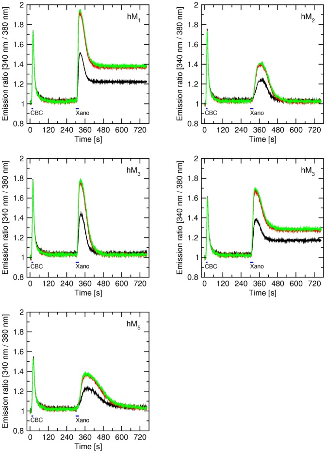
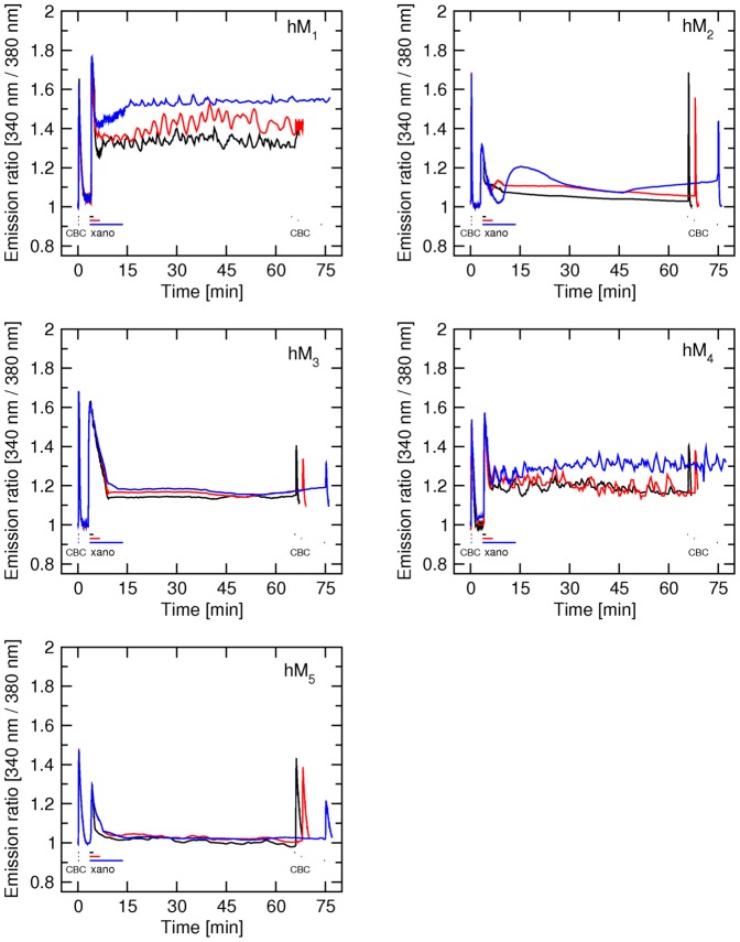
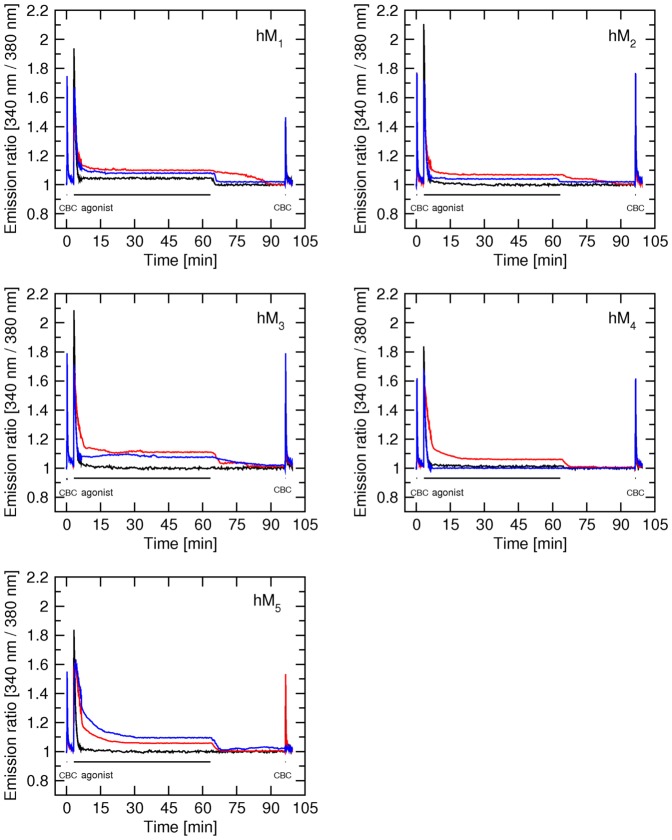
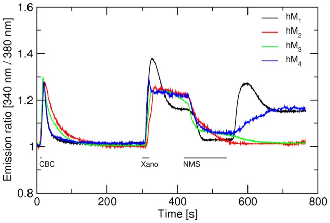
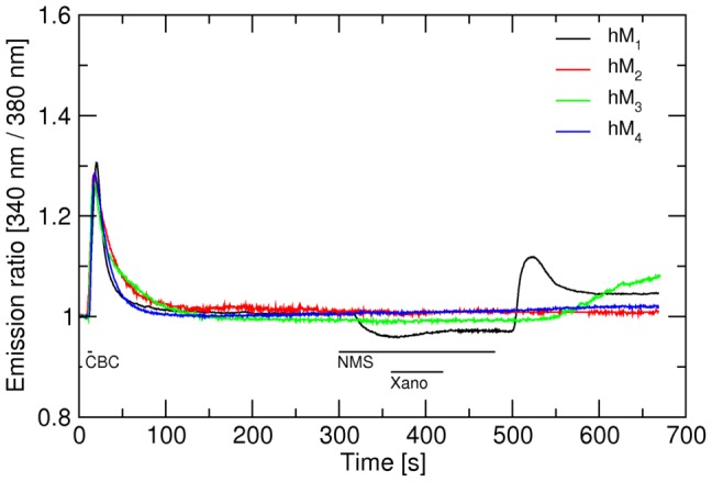
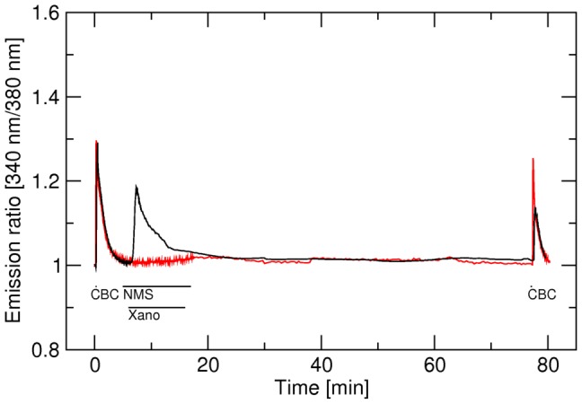
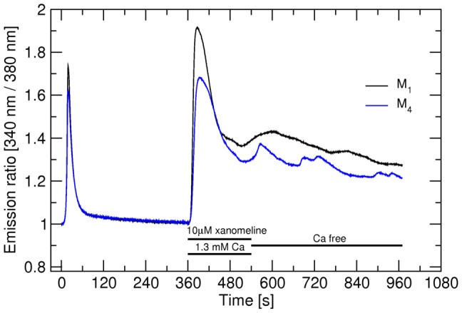
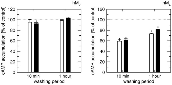
Xanomeline is an agonist endowed with functional preference for M1/M4 muscarinic acetylcholine receptors. It also exhibits both reversible and wash-resistant binding to and activation of these receptors. So far the mechanisms of xanomeline selectivity remain unknown. To address this question we employed microfluorometric measurements of intracellular calcium levels and radioligand binding to investigate differences in the short- and long-term effects of xanomeline among muscarinic receptors expressed individually in Chinese hamster ovary cells. 1/One-min exposure of cells to xanomeline markedly increased intracellular calcium at hM1 and hM4, and to a lesser extent at hM2 and hM3 muscarinic receptors for more than 1 hour. 2/Unlike the classic agonists carbachol, oxotremorine, and pilocarpine 10-min exposure to xanomeline did not cause internalization of any receptor subtype. 3/Wash-resistant xanomeline selectively prevented further increase in intracellular calcium by carbachol at hM1 and hM4 receptors. 4/After transient activation xanomeline behaved as a long-term antagonist at hM5 receptors. 5/The antagonist N-methylscopolamine (NMS) reversibly blocked activation of hM1 through hM4 receptors by xanomeline. 6/NMS prevented formation of xanomeline wash-resistant binding and activation at hM2 and hM4 receptors and slowed them at hM1, hM3 and hM5 receptors. Our results show commonalities of xanomeline reversible and wash-resistant binding and short-time activation among the five muscarinic receptor subtypes. However long-term receptor activation takes place in full only at hM1 and hM4 receptors. Moreover xanomeline displays higher efficacy at hM1 and hM4 receptors in primary phasic intracellular calcium release. These findings suggest the existence of particular activation mechanisms specific to these two receptors.


Conflict of interest statement

Competing Interests: The authors have declared that no competing interests exist.

Figures

Figure 1
Figure 1. Concentration response to acute treatment with xanomeline.
Cells were seeded, handled and loaded with Fura-2 as described in Methods. After an initial 10-s period cells were stimulated with 300 nM carbachol (CBC) for 5 s, washed with KHB for 5 min, then stimulated with 0.1 (black), 1 (red) or 10 µM (green) xanomeline (Xano) for 20 s and washed with KBH for 7 min. Traces are averages from 10 to 12 cells from representative experiment confirmed by 3 independent experiments. Signal variation (SD) among cells ranges from ±0.019 at the base line to ±0.035 at peaks. Parameters of calcium response are summarized in Table S2 in File S1. Calculated pEC50 and EMAX of response to xanomeline are in Table 1.
Figure 2
Figure 2. Effects of short-term application of xanomeline on the time-course of changes in intracellular calcium concentration in CHO cells expressing individual subtypes of muscarinic receptors.
The time-course of intracellular calcium concentration (abscissa) after stimulation of hM1 to hM5 muscarinic receptor subtypes with the agonists carbachol (CBC) and xanomeline was measured as described in Methods. First stimulation: After 10 s of initial (resting) period 300 nM carbachol was applied for 10 s and then washed. Second stimulation: Three min after the first stimulation 10 µM xanomeline was applied for 1 min (black curve), 3 min (red curve) or 10 min (blue curve) followed by washing. Third stimulation: One hour after the second stimulation 300 nM carbachol was applied for 10 s followed by washing. Intracellular calcium concentration (ordinate) is expressed as fluorescence intensity (340 nm/380 nm) ratio normalized to basal calcium level. Representative traces are averages of 8 to 12 best responding cells from one experiment. Signal variation (SD) among cells ranges from ±0.017 at the base line to ±0.063 at peaks. Results were confirmed in 5 additional independent experiments. Parameters of xanomeline effects are summarized in Table S4 in File S1.
Figure 3
Figure 3. Effects of long-term application of classic agonists on the time-course of changes in intracellular calcium concentration in CHO cells expressing individual subtypes of muscarinic receptors.
The time-course of changes in intracellular calcium concentration (abscissa) after stimulation of hM1 to hM5 muscarinic receptor subtypes with the agonists carbachol (CBC), oxotremorine and pilocarpine was measured as described in Methods. First stimulation: After 10 s of initial (resting) period 300 nM carbachol was applied for 10 s and then washed. Second stimulation: Three min after the first stimulation either 1 µM carbachol (black curve) or 1 µM oxotremorine (red curve) or 3 µM pilocarpine was applied for 1 hour followed by 30-min washing. Third stimulation: After washing following the second stimulation 300 nM carbachol was applied for 10 s followed by washing. Intracellular calcium concentration (ordinate) is expressed as fluorescence intensity (340 nm/380 nm) ratio normalized to basal calcium level. Representative traces are averages of 12 to 16 best responding cells from one experiment. Signal variation (SD) among cells ranges from ±0.018 at the base line to ±0.067 at peaks. Results were confirmed in 2 additional independent experiments. Parameters of agonist effects are summarized in Table S5 in File S1.
Figure 4
Figure 4. Effects of NMS on delayed elevation of intracellular calcium levels induced by short-term application of xanomeline at hM1 through hM4 receptors.
Changes in the concentration of intracellular calcium (ordinate) are expressed as changes in fluorescence intensity (340 nm/380 nm) ratio normalized to basal calcium level. First (control) stimulation: 300 nM carbachol (CBC) for 5 s was applied. Second stimulation: At 300 s stimulation with 10 µM xanomeline (Xano) was applied for 20 s. After 2-min washing with KHB cells were superfused with 10 µM NMS for 2 min and then washed with KHB for additional 4 min. Traces are averages of 8 to 12 best responding cells from one experiment. Signal variation (SD) among cells ranges from ±0.015 at the base line to ±0.037 at peaks. Results were confirmed in 5 additional independent experiments. Parameters of xanomeline effects are summarized in Table S6 in File S1.
Figure 5
Figure 5. Effects of NMS on formation of xanomeline wash-resistant activation at hM1 through hM4 receptors.
Changes in the concentration of intracellular calcium (ordinate) are expressed as changes in normalized fluorescence intensity (340 nm/380 nm) ratio normalized to basal calcium level. First (control) stimulation: Control 300 nM carbachol (CBC) was applied for 5 s. Second stimulation: At 5 min receptors were blocked by 10 µM of the antagonist NMS (1 min), then a mixture of 10 µM xanomeline (Xano) and 10 µM NMS was applied for 1 min and then 10 µM NMS was applied for an additional 1 min. Cells were then washed with KHB for additional 3 min. Representative traces are averages of 8 to 12 best responding cells from one experiment. Signal variation (SD) among cells ranges from ±0.015 at the base line to ±0.033 at peaks. Results were confirmed in 5 to 7 additional independent experiments. Parameters of xanomeline effects are summarized in Table S7 in File S1.
Figure 6
Figure 6. Effects of NMS on the formation of xanomeline wash-resistant action at hM5 receptors.
Changes in the concentration of intracellular calcium (ordinate) are expressed as changes in normalized fluorescence intensity (340 nm/380 nm) ratio normalized to basal calcium level. Red trace: First stimulation: Control 5-s stimulation with 300 nM carbachol (CBC) was performed. Second stimulation: At 5 min receptors were blocked by 10 µM of the antagonist NMS (1 min), then a mixture of 10 µM xanomeline (Xano) and 10 µM NMS was applied for 10 min and finally 10 µM NMS wash- applied for an additional 1 min. Third stimulation: After washing of the cells with KHB for 60-min 300 nM carbachol was applied for 5 s. Black trace: Control curve, same as red one but NMS was not applied. Representative traces are averages of 8 to 12 best responding cells from one experiment. Signal variation (SD) among cells ranges from ±0.015 at the base line to ±0.032 at peaks. Results were confirmed in 5 additional independent experiments.
Figure 7
Figure 7. Lack of effects of changing extracellular calcium on calcium oscillations.
Changes in the concentration of intracellular calcium (ordinate) are expressed as changes in normalized fluorescence intensity (340 nm/380 nm) ratio normalized to basal calcium level. Black trace: M1 receptors, blue trace: M4 receptors. First stimulation: Control 5-s stimulation with 300 nM carbachol (CBC) was performed. Second stimulation: After 6 min washing with KHB receptors were stimulated with 10 µM xanomeline for 3 min and then washed with calcium free KHB. Representative traces are averages of 12 best responding cells from one experiment. Signal variation (SD) among cells ranges from ±0.015 at the base line to ±0.063 at peaks. Results were confirmed in 2 additional independent experiments.
Figure 8
Figure 8. Effects of xanomeline on the accumulation of cyclic AMP.
Cells expressing hM2 (left) or hM4 (right) receptors were treated with 10 µM xanomeline for 3 min followed by washing for 10 min or 1 hour (coordinate) prior to 20-min incubation with 5 µM (white bars) or 20 µM (black bars) forskolin. Accumulation of [3H]cAMP (ordinate) is expressed as per cent of control accumulation of [3H]cAMP in xanomeline sham treated cells (corrected for content of protein). Data are means ± S.E.M. from 3 experiments performed in triplicates. *, different from control (sham treated) cells by t-test, P<0.05.

Similar articles

Cited by

References

    1. Bonner TI, Buckley NJ, Young AC, Brann MR (1987) Identification of a family of muscarinic acetylcholine receptor genes. Science 237: 527–532. - PubMed
    1. Jakubík J, El-Fakahany EE (2010) Allosteric Modulation of Muscarinic Acetylcholine Receptors. Pharmaceuticals 9: 2838–2860. - PMC - PubMed
    1. Christie JE, Shering A, Ferguson J, Glen AI (1981) Physostigmine and arecoline: effects of intravenous infusions in Alzheimer presenile dementia. Br J Psychiatry 138: 46–50. - PubMed
    1. Felder CC, Porter AC, Skillman TL, Zhang L, Bymaster FP, et al. (2001) Elucidating the role of muscarinic receptors in psychosis. Life Sci 68: 2605–2613. - PubMed
    1. Langmead CJ, Watson J, Reavill C (2008) Muscarinic acetylcholine receptors as CNS drug targets. Pharmacol Ther 117: 232–243. - PubMed

Publication types

MeSH terms

LinkOut - more resources