. 2014 Feb 18;9(2):e89150.
doi: 10.1371/journal.pone.0089150. eCollection 2014.

Fucoidan reduces secretion and expression of vascular endothelial growth factor in the retinal pigment epithelium and reduces angiogenesis in vitro

Affiliations

Fucoidan reduces secretion and expression of vascular endothelial growth factor in the retinal pigment epithelium and reduces angiogenesis in vitro

Michaela Dithmer et al. PLoS One. .

Abstract

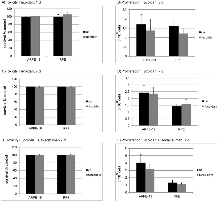
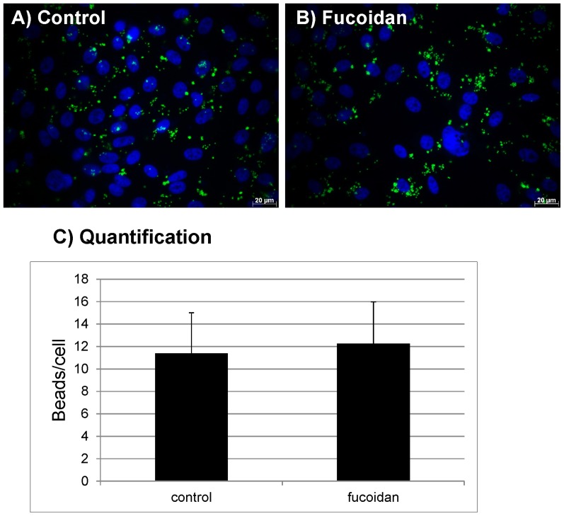
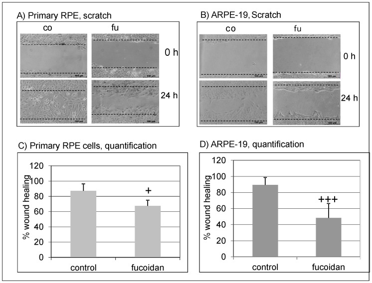
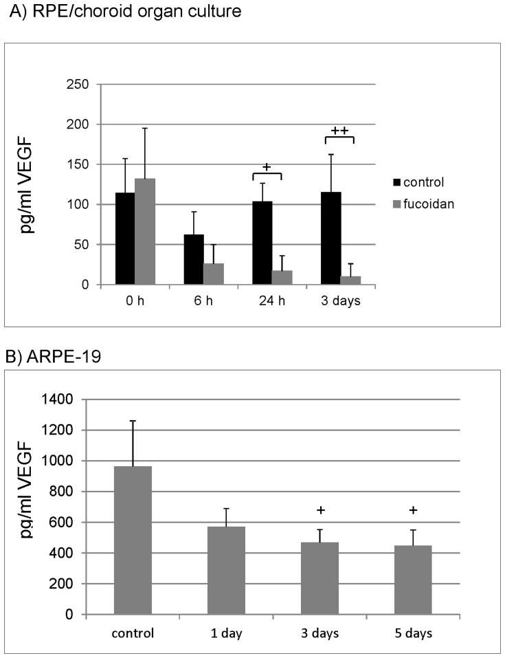
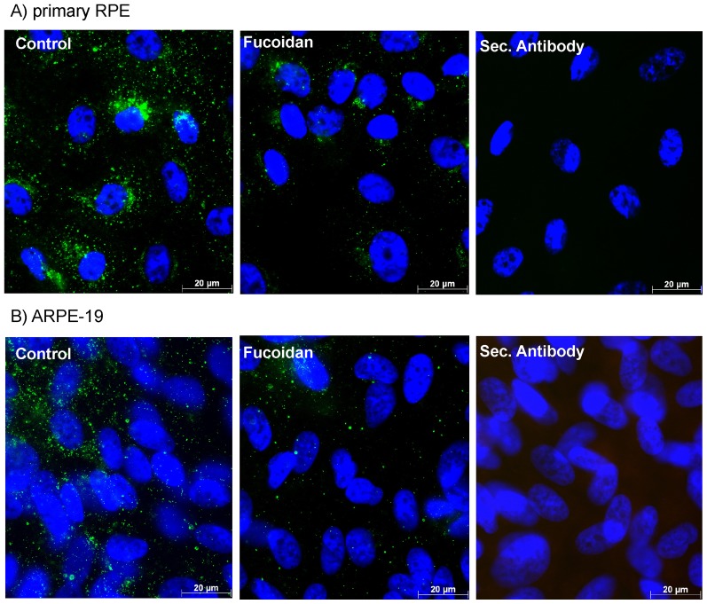
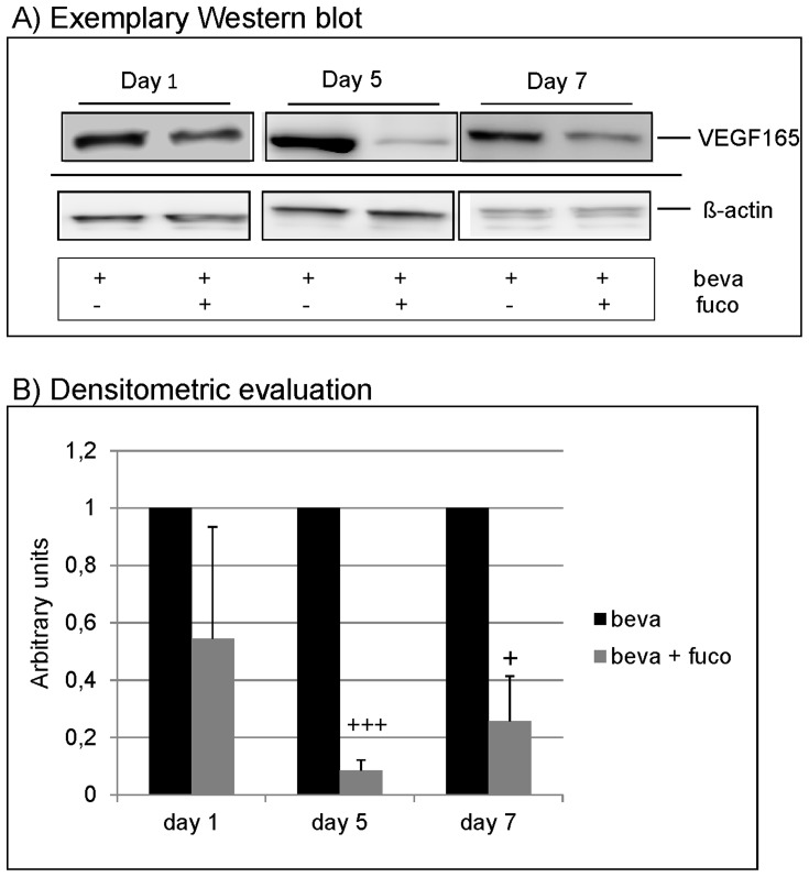
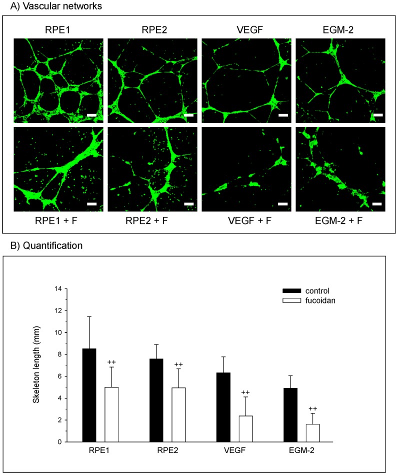
Fucoidan is a polysaccharide isolated from brown algae which is of current interest for anti-tumor therapy. In this study, we investigated the effect of fucoidan on the retinal pigment epithelium (RPE), looking at physiology, vascular endothelial growth factor (VEGF) secretion, and angiogenesis, thus investigating a potential use of fucoidan for the treatment of exudative age-related macular degeneration. For this study, human RPE cell line ARPE-19 and primary porcine RPE cells were used, as well as RPE/choroid perfusion organ cultures. The effect of fucoidan on RPE cells was investigated with methyl thiazolyl tetrazolium--assay, trypan blue exclusion assay, phagocytosis assay and a wound healing assay. VEGF expression was evaluated in immunocytochemistry and Western blot, VEGF secretion was evaluated in ELISA. The effect of fucoidan on angiogenesis was tested in a Matrigel assay using calcein-AM vital staining, evaluated by confocal laser scanning microcopy and quantitative image analysis. Fucoidan displays no toxicity and does not diminish proliferation or phagocytosis, but reduces wound healing in RPE cells. Fucoidan decreases VEGF secretion in RPE/choroid explants and RPE cells. Furthermore, it diminishes VEGF expression in RPE cells even when co-applied with bevacizumab. Furthermore, fucoidan reduces RPE-supernatant- and VEGF-induced angiogenesis of peripheral endothelial cells. In conclusion, fucoidan is a non-toxic agent that reduces VEGF expression and angiogenesis in vitro and may be of interest for further studies as a potential therapy against exudative age-related macular degeneration.


Conflict of interest statement

Competing Interests: Regarding the subject of this paper, the authors declare that no competing interests exist. Independent of this study, AK has been a consultant for and received lecture fees and research funding from Novartis. Harald Schmidt is the owner of MetaPhysiol and his affiliation with MetaPhysiol does not alter the authors' adherence to all the PLOS ONE policies on sharing data and materials.

Figures

Figure 1
Figure 1. Toxicity and proliferation.
To investigate toxicity, RPE or ARPE-19 cells were treated with 100 µg/ml fucoidan for 24 hours (A) or 7 days (C). In addition, cells were treated with a combination of fucoidan (100 µg/ml) and bevacizumab (250 µg/ml) for 7 days (E). Toxicity was measured with MTT test. Fucoidan did not exert toxic effects on RPE or ARPE-19 cells at any tested application (A,C,E). To investigate proliferation, a defined number of cells were seeded, cells were treated with fucoidan (100 µg/ml) and cell number was assessed after 3 days (B) and 7 days (D). In addition, cells were treated with a combination of fucoidan (100 µg/ml) and bevacizumab (250 µg/ml) and cell number was assessed after 7 days (F). No significant influence on proliferation was found. Significance was determined with student's t-test. Co  =  untreated control, fuco  =  fucoidan, beva  =  bevacizumab.
Figure 2
Figure 2. Phagocytosis.
Primary RPE cells were stimulated with 100 µg/ml fucoidan for 1 hour. RPE cells were exposed to FITC-labeled, photoreceptor outer segment opsonized beads for 4 hours and uptake of the beads was evaluated in fluorescence microscope. No influence of fucoidan on RPE phagocytosis was found. A) control, B) fucoidan, C) quantification of uptaken beads. Significance was determined with student's t-test.
Figure 3
Figure 3. Wound healing.
A wound was scratched in a confluent cell layer of primary porcine RPE cells and ARPE-19 cells. Cells were either untreated (control) or exposed to fucoidan (100 µg/ml) for 24 hours. Exemplary pictures of wound healing are depicted for primary RPE cells (A) and ARPE-19 cells (B). The percentage of coverage after 24 hours of wound healing is depicted in the graphs for primary RPE cells (C) and ARPE-19 cells (D). Fucoidan significantly reduces wound healing in both RPE and ARPE-19 cells. Significance was determined with student's t-test, + p<0.05; +++ p<0.001.
Figure 4
Figure 4. VEGF secretion.
VEGF secretion was investigated in RPE/choroid organ culture (A) and ARPE-19 cell culture (B). RPE/choroid perfusion organ cultures were treated with 100 µg/ml fucoidan for 3 days and supernatant was collected at 6 hours, 24 hours and 3 days for one hour. ARPE-19 cells were treated with 100 µg/ml fucoidan for five days, and medium was collected after 1 day, 3 days and 5 days. VEGF content was evaluated with ELISA. Fucoidan reduced VEGF content compared to control in organ culture after 24 hours and 3 days (A). In cell culture, a reduction of VEGF secretion can be found after 3 days and 5 days (B). Significance was determined with student's t-test, + p<0.05; ++ p<0.01.
Figure 5
Figure 5. VEGF165 expression.
Primary RPE cells (A) and ARPE-19 cells (B) were treated with 100 µg/ml fucoidan for 24 hours and the expression of intracellular VEGF (still containing signal peptide) was evaluated using immunocytochemistry. Cells treated with fucoidan exhibited a substantial decrease in intracellular VEGF expression.
Figure 6
Figure 6. VEGF expression in presence of bevacizumab.
ARPE-19 cells were treated with 250 µg/ml bevacizumab and 100 µg/ml fucoidan for 1 day, 5 days and 7 days. Controls cells were treated with bevacizumab only. Western blot for intracellular VEGF (still containing signal peptide) depicted a reduction of VEGF165 in Western blot at day 5 and day 7 (A), which was significant in densitrometric evaluation (B). Significance was determined with student's t-test, + p<0.05; +++ p<0.001. beva  =  bevacizumab, fuco  =  fucoidan.
Figure 7
Figure 7. Angiogenesis.
(A) Morphological appearance of OEC grown on Matrigel and stained with Calcein-AM which is converted to a green fluorescence by viable cells. Results indicated angiogenic structures in OEC treated with conditioned medium from RPE cells from different donors, VEGF and the EGM-2 (no VEGF). Additional treatment with fucoidan resulted in the reduction of vascular structures. (B) Quantitative image analysis depicting the skeleton length of angiogenic structures. Significance was determined with student's t-test, ++ p<0.01.

Similar articles

Cited by

References

    1. Schrader WF (2006) Age-related macular degeneration: a socioeconomic time bomb in our aging society. Ophthalmologe 103: 742–748. - PubMed
    1. Zarbin M (2004) Current concepts in the pathogenesis of age-related macular degeneration. Arch Ophthalmol 122: 598–614. - PubMed
    1. Sparrow JR, Ueda K, Zhou J (2012) Complement dysregulation in AMD: RPE-Bruch's membrane-choroid. Mol Aspects Med 33: 436–445. - PMC - PubMed
    1. Parmeggiani F, Romano MR, Costagliola C, Semeraro F, Incorvaia C, et al. (2012) Mechanism of inflammation in age-related macular degeneration. Mediators Inflamm 2012: 546786. - PMC - PubMed
    1. Miller DW, Joussen AM, Holz FG (2003) The molecular mechanisms of age-related macular degeneration. Ophthalmologe 100: 92–96. - PubMed

Publication types

MeSH terms