

Fibrous papule of the face, similar to tuberous sclerosis complex-associated angiofibroma, shows activation of the mammalian target of rapamycin pathway: evidence for a novel therapeutic strategy?
- PMID: 24558502
- PMCID: PMC3928451
- DOI: 10.1371/journal.pone.0089467
Fibrous papule of the face, similar to tuberous sclerosis complex-associated angiofibroma, shows activation of the mammalian target of rapamycin pathway: evidence for a novel therapeutic strategy?
Abstract
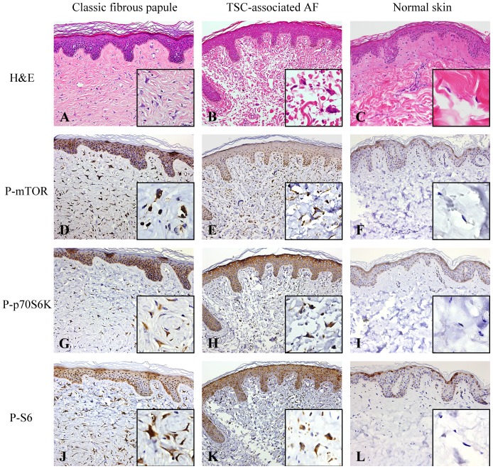
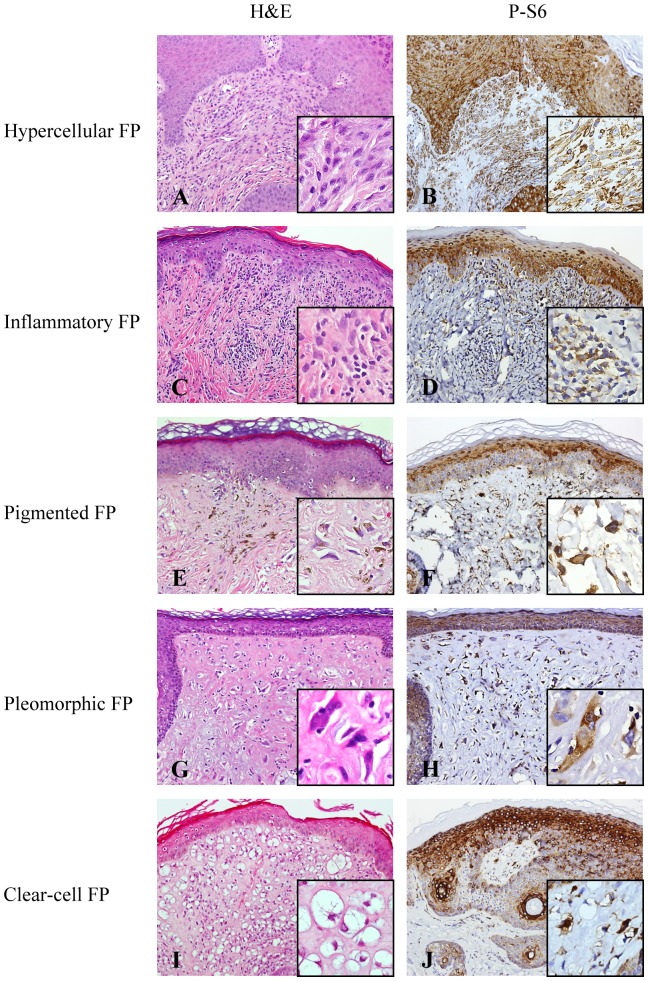
Fibrous papules of the face are hamartomas characterized by stellate-shaped stromal cells, multinucleated giant cells, and proliferative blood vessels in the dermis. The pathogenesis of fibrous papules remains unclear. There is a striking microscopic resemblance between fibrous papules and tuberous sclerosis complex (TSC)-associated angiofibromas. A germline mutation of the TSC1 or TSC2 gene, leading to activation of the mammalian target of rapamycin (mTOR) pathway, accounts for the pathogenesis of TSC-associated angiofibromas. Activated mTOR subsequently activates p70 ribosomal protein S6 kinase (p70S6K) and ribosomal protein S6 (S6) by phosphorylation. Rapamycin, a mTOR inhibitor, is effective in treating TSC-associated angiofibromas. The aim of this study was to understand whether the mTOR pathway is activated in fibrous papules. We studied immunoexpressions of phosphorylated (p-) mTOR effectors in fibrous papules, TSC-associated angiofibromas, and normal skin controls. P-mTOR, p-p70S6K and p-S6 were highly expressed in dermal stromal cells and epidermal keratinocytes in fibrous papules and TSC-associated angiofibromas but not in fibroblasts and epidermal keratinocytes of normal skin controls (p<0.001). The results suggest topical rapamycin may be a novel treatment option for fibrous papules.
Conflict of interest statement
Figures
Similar articles
-
Mesenchymal-epithelial interactions involving epiregulin in tuberous sclerosis complex hamartomas.Proc Natl Acad Sci U S A. 2008 Mar 4;105(9):3539-44. doi: 10.1073/pnas.0712397105. Epub 2008 Feb 21. Proc Natl Acad Sci U S A. 2008. PMID: 18292222 Free PMC article.
-
Pathogenesis of tuberous sclerosis subependymal giant cell astrocytomas: biallelic inactivation of TSC1 or TSC2 leads to mTOR activation.J Neuropathol Exp Neurol. 2004 Dec;63(12):1236-42. doi: 10.1093/jnen/63.12.1236. J Neuropathol Exp Neurol. 2004. PMID: 15624760
-
Loss of expression of tuberin and hamartin in tuberous sclerosis complex-associated but not in sporadic angiofibromas.J Cutan Pathol. 2003 Mar;30(3):174-7. doi: 10.1034/j.1600-0560.2003.2o066.x. J Cutan Pathol. 2003. PMID: 12641776
-
Rhebbing up mTOR: new insights on TSC1 and TSC2, and the pathogenesis of tuberous sclerosis.Cancer Biol Ther. 2003 Sep-Oct;2(5):471-6. doi: 10.4161/cbt.2.5.446. Cancer Biol Ther. 2003. PMID: 14614311 Review.
-
Pathogenesis of multifocal micronodular pneumocyte hyperplasia and lymphangioleiomyomatosis in tuberous sclerosis and association with tuberous sclerosis genes TSC1 and TSC2.Pathol Int. 2001 Aug;51(8):585-94. doi: 10.1046/j.1440-1827.2001.01242.x. Pathol Int. 2001. PMID: 11564212 Review.
Cited by
-
Is mTOR Inhibitor Good Enough for Treatment All Tumors in TSC Patients?J Cancer. 2016 Jul 21;7(12):1621-1631. doi: 10.7150/jca.14747. eCollection 2016. J Cancer. 2016. PMID: 27698899 Free PMC article. Review.
-
Clinical and genetic analysis of tuberous sclerosis complex-associated renal angiomyolipoma in Chinese pedigrees.Oncol Lett. 2017 Dec;14(6):7085-7090. doi: 10.3892/ol.2017.7079. Epub 2017 Sep 27. Oncol Lett. 2017. PMID: 29344138 Free PMC article.
-
Benign tumors in TSC are amenable to treatment by GD3 CAR T cells in mice.JCI Insight. 2021 Nov 22;6(22):e152014. doi: 10.1172/jci.insight.152014. JCI Insight. 2021. PMID: 34806651 Free PMC article.
References
-
- Graham JH, Sanders JB, Johnson WC, Helwig EB (1965) Fibrous papule of the nose: a clinicopathological study. J Invest Dermatol 45: 194–203. - PubMed
-
- Bansal C, Stewart D, Li A, Cockerell CJ (2005) Histologic variants of fibrous papule. J Cutan Pathol 32: 424–428. - PubMed
-
- Meigel WN, Ackerman AB (1979) Fibrous papule of the face. Am J Dermatopathol 4: 329–340. - PubMed
-
- Guitart J, Bergfeld WF, Tuthill RJ (1991) Fibrous papule of the nose with granular cells: two cases. J Cutan Pathol 18: 284–287. - PubMed
-
- Ragaz A, Berezowsky V (1979) Fibrous papule of the face. A study of five cases by electron microscopy. Am J Dermatopathol 1: 353–356. - PubMed
Publication types
MeSH terms
Substances
LinkOut - more resources
Full Text Sources
Other Literature Sources
Medical
Miscellaneous
