

Prevalence of Talon cusp in Indian population
- PMID: 24558520
- PMCID: PMC3908805
- DOI: 10.4317/jced.50650
Prevalence of Talon cusp in Indian population
Abstract
Aim: To investigate the prevalence of the talon cusps in a sample of Indian dental patients and their distribution among different types of teeth. To determine the presence of other dental anomalies associated with the talon cusps.
Method: 2740 out patients (1523 males and 1217 females) attending Oral Medicine department from November 2010 to January 2011 were screened for the presence of talon cusps and were subjected to Intra Oral Peri-apical (IOPA) radiograph to rule out any associated anomalies or peri-apical changes.
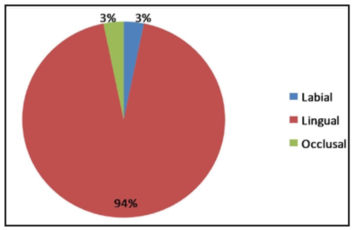
Results: Talon cusps were detected in 16 out of 2740 patients (person prevalence 0.58%). Thirty one teeth were found to have talon cusp. Maxillary lateral incisors were the most commonly affected teeth (54.8%, 17 teeth), followed by maxillary central incisors and canines (16.12%, 5 teeth).Talon cusp was found in two mandibular central incisors (6.45%) and one each in mandibular second and third molar (3.22% each). Seventeen teeth in 7 patients (54.83%) were found to be associated with anomalies like dens invagination (6 teeth, 19.35%), impacted 13, 23 (6 teeth, 19.35%), partial anodontia (3 teeth, 9.67%), geographic and fissured tongue (2 teeth, 6.45%). Peri-apical granuloma was found in one tooth with talon cusp associated with dens invaginatus. None of the patients were found to be associated with any syndromes.
Conclusion: Attention should be paid to the presence of the talon cusp and the associated anomalies. Early diagnosis of the talon cusp can help the clinician in preventing the further complications. Key words:Orthopantomography, atheroma, stroke.
Figures


Similar articles
-
Prevalence and characteristics of talon cusps in Turkish population.Dent Res J (Isfahan). 2016 Mar-Apr;13(2):145-50. doi: 10.4103/1735-3327.178200. Dent Res J (Isfahan). 2016. PMID: 27076829 Free PMC article.
-
Survey of talon cusps in the permanent dentition of a Turkish population.J Contemp Dent Pract. 2008 Jul 1;9(5):84-91. J Contemp Dent Pract. 2008. PMID: 18633473
-
The characteristics and occurrence of the talon cusps in Turkish population: a retrospective sample study.Surg Radiol Anat. 2016 Nov;38(9):1105-1110. doi: 10.1007/s00276-016-1653-6. Epub 2016 Feb 22. Surg Radiol Anat. 2016. PMID: 26899859
-
Talon cusps occurring concurrently with dens invaginatus on a permanent maxillary lateral incisor: a case report and literature review.Gen Dent. 2014 May-Jun;62(3):e14-8. Gen Dent. 2014. PMID: 24784523 Review.
-
Mandibular talon cusps: A Systematic review and data analysis.J Clin Exp Dent. 2014 Oct 1;6(4):e408-13. doi: 10.4317/jced.51476. eCollection 2014 Oct. J Clin Exp Dent. 2014. PMID: 25593665 Free PMC article. Review.
Cited by
-
Dens Invaginatus: A Comprehensive Review of Classification and Clinical Approaches.Medicina (Kaunas). 2025 Jul 16;61(7):1281. doi: 10.3390/medicina61071281. Medicina (Kaunas). 2025. PMID: 40731909 Free PMC article. Review.
-
Prevalence and characteristics of talon cusps in Turkish population.Dent Res J (Isfahan). 2016 Mar-Apr;13(2):145-50. doi: 10.4103/1735-3327.178200. Dent Res J (Isfahan). 2016. PMID: 27076829 Free PMC article.
-
Facial Cellulitis Due to Type I Talon Cusp in a Pediatric Patient: A Case Report.Cureus. 2023 Jan 20;15(1):e34011. doi: 10.7759/cureus.34011. eCollection 2023 Jan. Cureus. 2023. PMID: 36814740 Free PMC article.
-
Double facial talons on maxillary incisor-A rare case report and new proposed classification system.J Oral Maxillofac Pathol. 2022 Jul-Sep;26(3):423. doi: 10.4103/jomfp.jomfp_43_22. Epub 2022 Oct 17. J Oral Maxillofac Pathol. 2022. PMID: 36588837 Free PMC article.
-
Evaluating the Prevalence and Distribution of Dental Anomalies in the Permanent Dentition of Patients Seeking Dental Care.Cureus. 2022 Oct 10;14(10):e30156. doi: 10.7759/cureus.30156. eCollection 2022 Oct. Cureus. 2022. PMID: 36397922 Free PMC article.
References
-
- Mellor JK, Ripa LW. Talon cusp: a clinically significant anomaly. Oral Surg Oral Med Oral Pathol. 1970;29:225–8. - PubMed
-
- Lee CK, King NM, Lo EC, Cho SY. The relationship between a primary maxillary incisor with a talon cusp and the permanent successor: a study of 57 cases. Int J Paediatr Dent. 2007;17:178–85. - PubMed
-
- Dankner E, Harari D, Rotstein I. Dens evaginatus of anterior teeth. Literature review and radiographic survey of 15,000 teeth. Oral Surg Oral Med Oral Pathol Oral Radiol Endod. 1996;81:472–5. - PubMed
-
- Lomcali G, Hazar S, Altinbulak H. Talon cusp: report of five cases. Quintessence Int. 1994;25:431–3. - PubMed
-
- Rantanen AV. Talon cusp. Oral Surg Oral Med Oral Pathol. 1971;32:398–400. - PubMed
LinkOut - more resources
Full Text Sources