Orbital decompression in thyroid eye disease
- PMID: 24558591
- PMCID: PMC3914264
- DOI: 10.5402/2012/739236
Orbital decompression in thyroid eye disease
Abstract
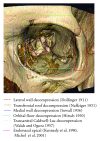
Though enlargement of the bony orbit by orbital decompression surgery has been known for about a century, surgical techniques vary all around the world mostly depending on the patient's clinical presentation but also on the institutional habits or the surgeon's skills. Ideally every surgical intervention should be tailored to the patient's specific needs. Therefore the aim of this paper is to review outcomes, hints, trends, and perspectives in orbital decompression surgery in thyroid eye disease regarding different surgical techniques.
Figures


References
-
- Meyer DR. Surgical management of Graves orbitopathy. Current Opinion in Ophthalmology. 1999;10(5):343–351. - PubMed
-
- Bartley GB, Fatourechi V, Kadrmas EF, et al. The treatment of Graves’ ophthalmopathy in an incidence cohort. American Journal of Ophthalmology. 1996;121(2):200–206. - PubMed
-
- Wiersinga WM, Prummel MF. Graves’ ophthalmopathy: a rational approach to treatment. Trends in Endocrinology and Metabolism. 2002;13(7):280–287. - PubMed
-
- Bartalena L, Baldeschi L, Dickinson AJ, et al. Consensus statement of the European group on Graves' orbitopathy (EUGOGO) on management of Graves' orbitopathy. Thyroid. 2008;18(3):333–346. - PubMed
-
- Char D. Thyroid Eye Disease. 3rd edition. Boston, Mass, USA: Butterworth-Heinemann; 1997.
Publication types
LinkOut - more resources
Full Text Sources
