. 2014 Mar 25;8(3):2148-60.
doi: 10.1021/nn405033r. Epub 2014 Feb 27.

A biodegradable nanoparticle platform for the induction of antigen-specific immune tolerance for treatment of autoimmune disease

Affiliations

A biodegradable nanoparticle platform for the induction of antigen-specific immune tolerance for treatment of autoimmune disease

Zoe Hunter et al. ACS Nano. .

Abstract

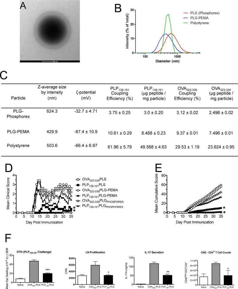
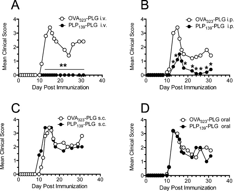
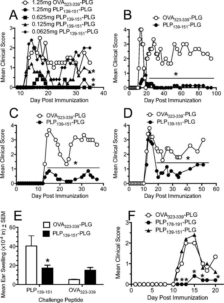
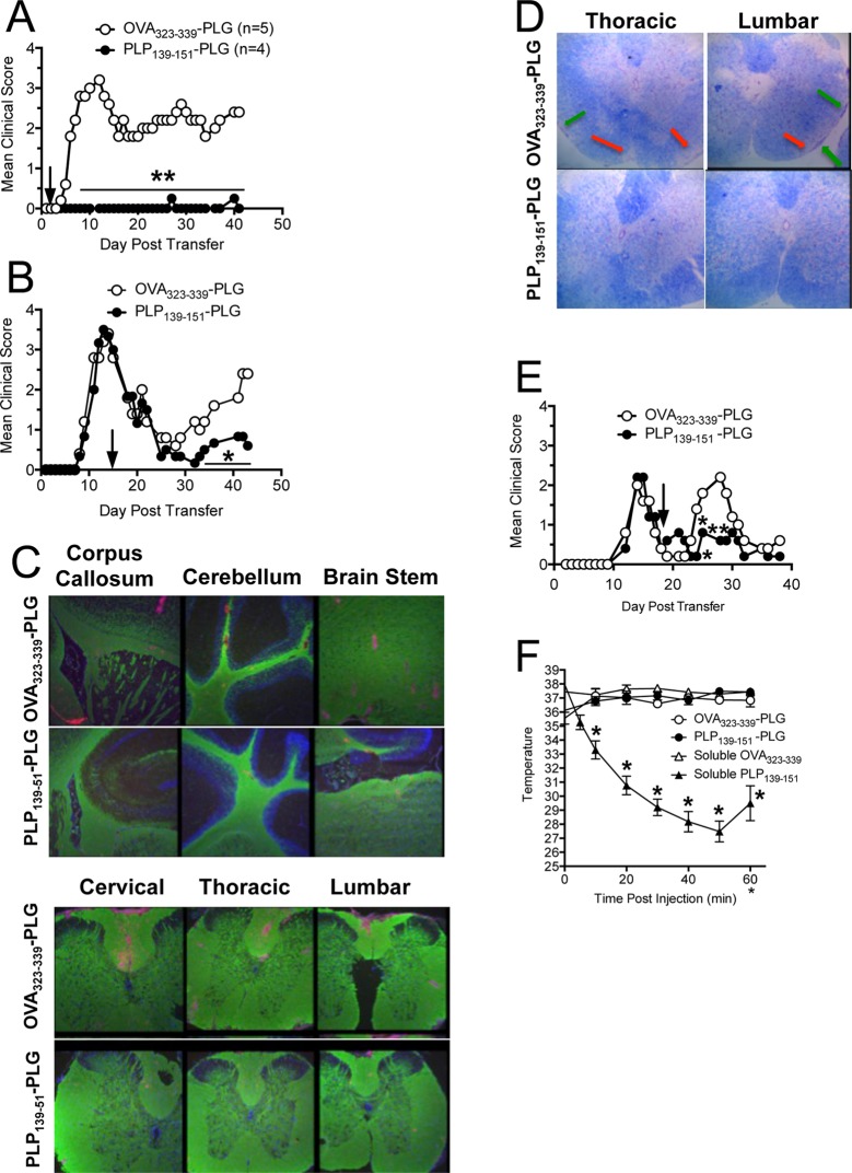
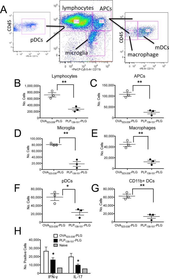
Targeted immune tolerance is a coveted therapy for the treatment of a variety of autoimmune diseases, as current treatment options often involve nonspecific immunosuppression. Intravenous (iv) infusion of apoptotic syngeneic splenocytes linked with peptide or protein autoantigens using ethylene carbodiimide (ECDI) has been demonstrated to be an effective method for inducing peripheral, antigen-specific tolerance for treatment of autoimmune disease. Here, we show the ability of biodegradable poly(lactic-co-glycolic acid) (PLG) nanoparticles to function as a safe, cost-effective, and highly efficient alternative to cellular carriers for the induction of antigen-specific T cell tolerance. We describe the formulation of tolerogenic PLG particles and demonstrate that administration of myelin antigen-coupled particles both prevented and treated relapsing-remitting experimental autoimmune encephalomyelitis (R-EAE), a CD4 T cell-mediated mouse model of multiple sclerosis (MS). PLG particles made on-site with surfactant modifications surpass the efficacy of commercially available particles in their ability to couple peptide and to prevent disease induction. Most importantly, myelin antigen-coupled PLG nanoparticles are able to significantly ameliorate ongoing disease and subsequent relapses when administered at onset or at peak of acute disease, and minimize epitope spreading when administered during disease remission. Therapeutic treatment results in significantly reduced CNS infiltration of encephalitogenic Th1 (IFN-γ) and Th17 (IL-17a) cells as well as inflammatory monocytes/macrophages. Together, these data describe a platform for antigen display that is safe, low-cost, and highly effective at inducing antigen-specific T cell tolerance. The development of such a platform carries broad implications for the treatment of a variety of immune-mediated diseases.


Figures

Figure 1
Figure 1
Peptide-coupled PLG-PEMA nanoparticles are efficient carriers for induction of antigen-specific tolerance for prevention of EAE. (A) Micrograph of PLG-PEMA particles. (B and C) Size distribution, average size (nm), ζ-potential (mV), peptide coupling efficiency (%), and amount of OVA323–339 and PLP139–151 peptides coupled per milligram of PLG-PEMA particles prepared in the laboratory in comparison to PLG particles purchased from Phosphorex (PLGPHOSPHOREX) and polystyrene (PS) particles purchased from Polysciences. (D and E) Groups of 6–8 week old SJL/J mice were injected intravenously (iv) with 1.25 mg of the various carboxylated nanoparticles coupled to OVA323–339 or PLP139–151 7d prior to induction of EAE by subcutaneous (sc) immunization with PLP139–151/CFA. Disease symptoms were scored by daily assessment of mean clinical score (D) as well as the mean cumulative clinical score (E) for the next 35 days. All experimental groups consisted of 5–7 mice and are representative of three separate experiments. Mean clinical scores and mean cumulative clinical scores for the mice tolerized with PLP139–151 coupled to either PLG-PEMA or PS were significantly less than scores for mice treated with the appropriate OVA323–339 coupled control particles (*p ≤ 0.05, ANOVA). (F) At day +14, 24 h DTH responses to ear challenge with 10 μg of PLP139–151 were determined in 4–5 selected mice from the PLG-PEMA treated groups, and proliferative and IL-17 responses of draining lymph node T cells were determined following 72 h in vitro stimulation with peptide. At the end of the clinical observation period, the numbers of CNS-infiltrating T cells were enumerated in 3–4 selected mice from each group. Responses in PLP139–151-PLG treated mice were significantly less than those in OVA323–339-PLG treated controls (*p ≤ 0.05, **p ≤ 0.01, ***p ≤ 0.001, ANOVA).
Figure 2
Figure 2
Efficient induction of tolerance with peptide-coupled PLG-PEMA nanoparticles is dependent on route of administration. The 6–8 week old female SJL/J mice were treated with 1.25 mg of PLG-PEMA nanoparticles coupled with OVA323–339 or PLP139–151via intravenous (iv) (A), intraperitoneal (ip) (B), subcutaneous (sc) (C), or oral (D) routes of administration 7d prior to induction of EAE by sc immunization with PLP139–151/CFA. Disease symptoms were scored by daily assessment of mean clinical scores for the next 32 days. Data represent one of two representative experiments. Mean clinical scores were significantly less for mice treated with PLP139–151-PLG than for mice treated with the OVA323–339-coupled control PLG particles (*p ≤ 0.05, **p ≤ 0.01 ANOVA).
Figure 3
Figure 3
Preventative treatment with myelin antigen-coupled PLG-PEMA nanoparticles induces long-term, antigen-specific tolerance. (A) Optimal dosing of Ag-PLG-PEMA for preventative tolerance was determined by iv administration of increasing amounts of PLP139–151-PLG to SJL/J mice 7 days prior to disease induction by sc immunization with PLP139–151/CFA. Mice were monitored for development of clinical disease for 35 days after priming. (B–D) Duration of tolerance in mice was determined by iv administration of 1.25 mg of PLP139–151- or OVA323–339-PLG nanoparticles to 6–8 week old naïve female SJL/J mice which were primed sc with PLP139–151/CFA 7 days (B), 25 days (C), or 50 days (D) later. Mice were scored for development of clinical disease for the indicated time periods. (E) On day 8 relative to PLP139–151/CFA priming, DTH responses, as measured by 24 h swelling responses induced by sc ear challenge with PLP139–151 or OVA323–339 control peptide, in representative animals from panel B were determined. (F) The 6–8 week old SJL/J mice were treated iv with 1.25 mg of OVA323–339-PLG, PLP178–191-PLG, or PLP139–151-PLG 7d prior to sensitization with PLP178–191/CFA, and disease was monitored for 35 days thereafter. All experimental groups consisted of 5–7 mice and are representative of 2–3 repeats. Differences in mean clinical scores and DTH responses were significantly less than the responses in groups tolerized with the irrelevant OVA323–339-PLG nanoparticles (*p ≤ 0.05, ANOVA).
Figure 4
Figure 4
Myelin Ag-coupled PLG-PEMA nanoparticles safely and effectively treat established EAE and prevent disease relapses. EAE was induced in 6–8 week old female SJL/J mice by adoptive transfer of 2.5 × 10⁶ PLP139–151-specific blasts. Mice were injected iv with either 1.25 mg of OVA323–339- or PLP139–151-PLG-PEMA nanoparticles 2 days (A) or 14 days (B) following cell transfer and mice were followed for clinical disease for the indicated number of days. (C) Brain and spinal cords were collected from the mice in panel A tolerized with OVA323–339- or PLP139–151-PLG on day +2 for histological analysis on day 42, and sections were stained for PLP protein (green) and CD45 (red). (D) Spinal cord sections from mice from panel B were stained with Luxol Fast Blue. Areas of demyelination are indicated by red arrows; areas of cellular infiltration are indicated by green arrows. Higher magnification images are shown in Figure S1. (E) To test the efficacy of Ag-PLG nanoparticles to treat EAE mice during disease remission for prevention of disease relapses, SJL/J mice with EAE induced by sc priming with PLP178–191/CFA were tolerized by iv infusion of OVA323–339- or PLP139–151-PLG on day +18, and clinical disease was scored for an additional 21 days. (F) To determine the potential of Ag-PLG tolerance to trigger anaphylaxis in primed mice, EAE was induced in 6–8 week old female SJL/J mice by subcutaneous injection of PLP139–151/CFA and the mice were treated by iv infusion of 200 μg of soluble OVA323–339 or PLP139–151, or the same peptides coupled to PLG-PEMA nanoparticles on day +21. Temperature of the injected animals was monitored and recorded every 10 min for 1 h following injection as a measure of induction of anaphylaxis. All experimental groups consisted of 5–7 mice and are representative of 2–3 separate experiments. Mean clinical scores were significantly less for mice treated with PLP139–151-PLG than for mice treated with the OVA323–339-coupled control PLG particles (*p ≤ 0.05, **p ≤ 0.01 ANOVA).
Figure 5
Figure 5
Induction of therapeutic tolerance with myelin Ag-coupled PLG-PEMA nanoparticles reduces CNS immune infiltration and cytokine secretion. (A) SJL/J mice were injected iv with PLG-PEMA nanoparticles coupled with OVA323–339 or PLP139–151 2d following EAE induction by adoptive transfer of activated PLP139–151-specfic T cell blasts. At the peak of disease in the OVA323–339-PLG treated controls (d+14), brains and spinal cords were removed and processed into single cell suspensions, and the numbers of infiltrating immune cells were analyzed by flow cytometry using the indicated gating scheme. The numbers of lymphocytes (B), APCs (C), microglia (D), macrophages (E), myeloid dendritic cells (mDCs) (F), and conventional peripheral dendritic cells (pDCs) (G) were enumerated by flow cytometry. (H) CNS cell preparations from the two treated groups as well as from naïve controls were stimulated with PMA and ionomycin for 5 h prior to intracellular staining for IFN-γ and IL-17A. Representative dot plots are shown in Figure S2. Data is representative of 2–3 separate experiments. Numbers of the various cell subsets infiltrating the CNS were significantly less for mice treated with PLP139–151-PLG than for mice treated with the OVA323–339-coupled control PLG particles (*p ≤ 0.05, **p ≤ 0.01 ANOVA).

References

    1. American Autoimmune Related Diseases Association The Cost Burden of Autoimmune Disease: The Latest Front in the War on Healthcare Spending; AARDA: Eastpointe, MI, 2011. http://www.aarda.org/pdf/cbad.pdf.
    1. Chatenoud L.; Thervet E.; Primo J.; Bach J. F. Anti-CD3 Antibody Induces Long-Term Remission of Overt Autoimmunity in Nonobese Diabetic Mice. Proc. Natl. Acad. Sci. U.S.A. 1994, 91, 123–127. - PMC - PubMed
    1. Chatenoud L.; Primo J.; Bach J. F. CD3 Antibody-Induced Dominant Self Tolerance in Overtly Diabetic Nod Mice. J. Immunol. 1997, 158, 2947–2954. - PubMed
    1. Belghith M.; Bluestone J. A.; Barriot S.; Megret J.; Bach J. F.; Chatenoud L. Tgf-Beta-Dependent Mechanisms Mediate Restoration of Self-Tolerance Induced by Antibodies to CD3 in Overt Autoimmune Diabetes. Nat. Med. 2003, 9, 1202–1208. - PubMed
    1. Kohm A. P.; Williams J. S.; Bickford A. L.; McMahon J. S.; Chatenoud L.; Bach J. F.; Bluestone J. A.; Miller S. D. Treatment with Nonmitogenic Anti-CD3Monoclonal Antibody Induces CD4+ T Cell Unresponsiveness and Functional Reversal of Established Experimental Autoimmune Encephalomyelitis. J. Immunol. 2005, 174, 4525–4534. - PubMed

Publication types

MeSH terms