

Matricellular protein thrombospondins: influence on ocular angiogenesis, wound healing and immuneregulation
- PMID: 24559320
- PMCID: PMC4278647
- DOI: 10.3109/02713683.2013.877936
Matricellular protein thrombospondins: influence on ocular angiogenesis, wound healing and immuneregulation
Abstract
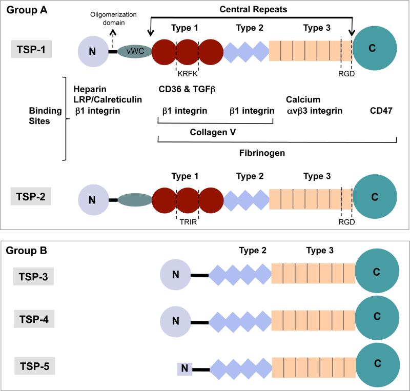
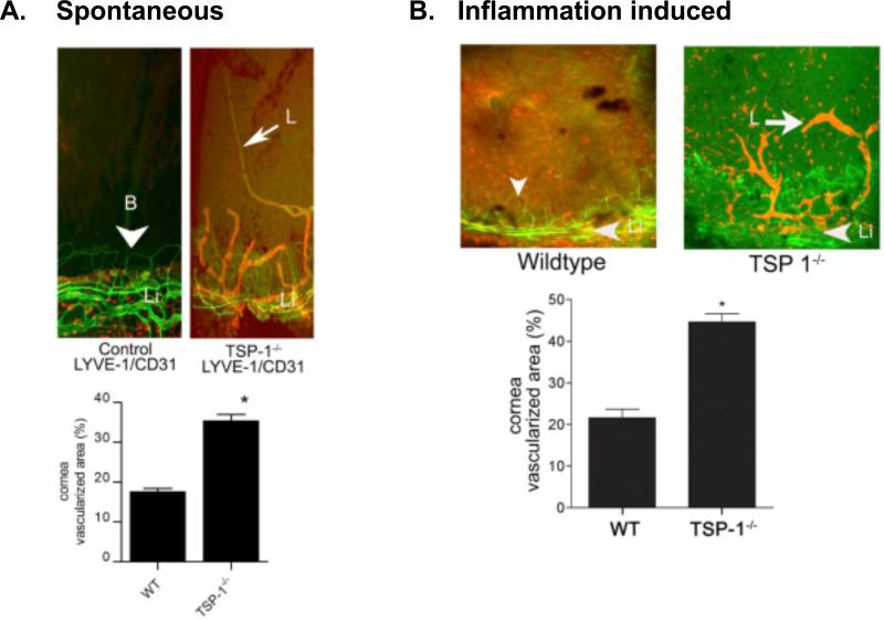
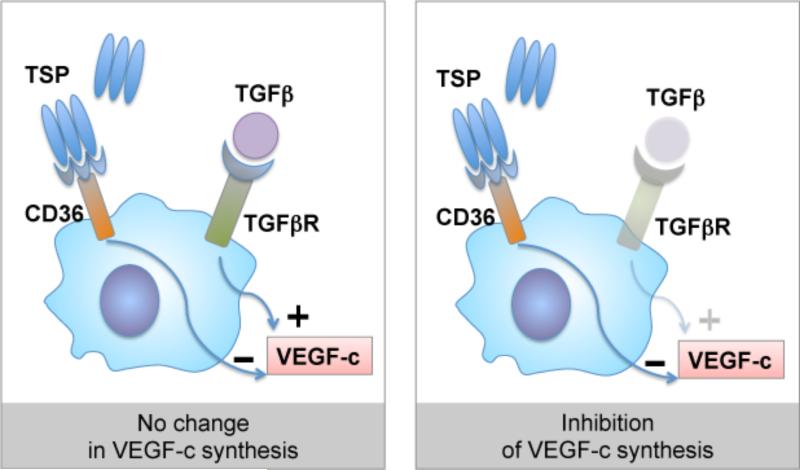
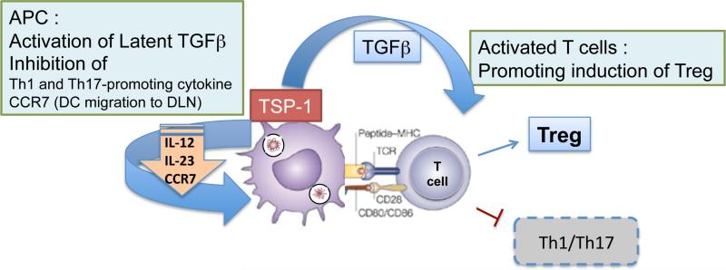
Thrombospondins are a family of large multi-domain glycoproteins described as matricelluar proteins based on their ability to interact with a broad range of receptors, matrix molecules, growth factors or proteases, and to modulate array of cellular functions including intracellular signaling, proliferation and migration. Two members of the thrombospondin family, thrombospondin 1 (TSP-1) and thrombospondin 2 (TSP-2) are studied extensively to determine their structure and function. While expressed at low levels in normal adult tissues, their increased expression is seen predominantly in response to cellular perturbations. Despite structural similarities, a notable functional difference between TSP-1 and TSP-2 includes the ability of former to activate of latent TGF-β and its competitive inhibition by the latter. Both these thrombospondins are reported to play important roles in TGF-β rich ocular environment with most reports related to TSP-1. They are expressed by many ocular cell types and detectable in the aqueous and vitreous humor. TSP-1 and TSP-2 influence many cellular interactions in the eye such as angiogenesis, cell migration, wound healing, TGF-β activation and regulation of inflammatory immune responses. Together, these processes are known to contribute to the immune privilege status of the eye. Emerging roles of TSP-1 and TSP-2 in ocular functions and pathology are reviewed here.
Keywords: Angiogenesis; immuneregulation; inflammation; lymphangiogenesis; wound healing.
Figures
Similar articles
-
Thrombospondin-1 as a Regulator of Corneal Inflammation and Lymphangiogenesis: Effects on Dry Eye Disease and Corneal Graft Immunology.J Ocul Pharmacol Ther. 2015 Sep;31(7):376-85. doi: 10.1089/jop.2015.0020. Epub 2015 Jul 8. J Ocul Pharmacol Ther. 2015. PMID: 26154823 Review.
-
The Roles of Thrombospondins in Hemorrhagic Stroke.Biomed Res Int. 2017;2017:8403184. doi: 10.1155/2017/8403184. Epub 2017 Oct 30. Biomed Res Int. 2017. PMID: 29214179 Free PMC article. Review.
-
Thrombospondin-4 mediates TGF-β-induced angiogenesis.Oncogene. 2017 Sep 7;36(36):5189-5198. doi: 10.1038/onc.2017.140. Epub 2017 May 8. Oncogene. 2017. PMID: 28481870 Free PMC article.
-
CD36-TSP-HRGP interactions in the regulation of angiogenesis.Curr Pharm Des. 2007;13(35):3559-67. doi: 10.2174/138161207782794185. Curr Pharm Des. 2007. PMID: 18220792 Review.
-
The role of the thrombospondins in healing myocardial infarcts.Cardiovasc Hematol Agents Med Chem. 2007 Jan;5(1):21-7. doi: 10.2174/187152507779315813. Cardiovasc Hematol Agents Med Chem. 2007. PMID: 17266545 Review.
Cited by
-
Vitamin D Receptor Expression Limits the Angiogenic and Inflammatory Properties of Retinal Endothelial Cells.Cells. 2023 Jan 16;12(2):335. doi: 10.3390/cells12020335. Cells. 2023. PMID: 36672270 Free PMC article.
-
Initiation of fibrosis in the integrin Αvβ6 knockout mice.Exp Eye Res. 2019 Mar;180:23-28. doi: 10.1016/j.exer.2018.11.027. Epub 2018 Nov 28. Exp Eye Res. 2019. PMID: 30500364 Free PMC article.
-
Phase 1 dose expansion and biomarker study assessing first-in-class tumor microenvironment modulator VT1021 in patients with advanced solid tumors.Commun Med (Lond). 2024 May 21;4(1):95. doi: 10.1038/s43856-024-00520-z. Commun Med (Lond). 2024. PMID: 38773224 Free PMC article.
-
Expression of pigment epithelium-derived factor and thrombospondin-1 regulate proliferation and migration of retinal pigment epithelial cells.Physiol Rep. 2015 Jan 19;3(1):e12266. doi: 10.14814/phy2.12266. Print 2015 Jan 1. Physiol Rep. 2015. PMID: 25602019 Free PMC article.
-
The immunoregulatory role of corneal epithelium-derived thrombospondin-1 in dry eye disease.Ocul Surf. 2018 Oct;16(4):470-477. doi: 10.1016/j.jtos.2018.07.005. Epub 2018 Jul 25. Ocul Surf. 2018. PMID: 30055331 Free PMC article.
References
-
- Ide M, Ishii H, Mukae H, Iwata A, Sakamoto N, Kadota J, et al. High serum levels of thrombospondin-1 in patients with idiopathic interstitial pneumonia. Respir Med. 2008;102(11):1625–30. - PubMed
Publication types
MeSH terms
Substances
Grants and funding
LinkOut - more resources
Full Text Sources
Other Literature Sources
Medical
Miscellaneous