Prospects for lentiviral vector mediated prostaglandin F synthase gene delivery in monkey eyes in vivo
- PMID: 24559478
- PMCID: PMC4134385
- DOI: 10.3109/02713683.2014.884593
Prospects for lentiviral vector mediated prostaglandin F synthase gene delivery in monkey eyes in vivo
Abstract
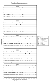
Currently, the most effective outflow drugs approved for clinical use are prostaglandin F2α analogues, but these require daily topical self-dosing and have various intraocular, ocular surface and extraocular side effects. Lentiviral vector-mediated delivery of the prostaglandin F synthase (PGFS) gene, resulting in long-term reduction of intraocular pressure (IOP), may eliminate off-target tissue effects and the need for daily topical PGF2α self-administration. Lentiviral vector-mediated delivery of the PGFS gene to the anterior segment has been achieved in cats and non-human primates. Although these results are encouraging, our studies have identified a number of challenges that need to be overcome for prostaglandin gene therapy to be translated into the clinic. Using examples from our work in non-human primates, where we were able to achieve a significant reduction in IOP (2 mm Hg) for 5 months after delivery of the cDNA for bovine PGF synthase, we identify and discuss these issues and consider several possible solutions.
Keywords: Glaucoma; gene therapy; lentivirus; prostaglandin synthase; trabecular meshwork.
Figures


Similar articles
-
Gene transfer to the outflow tract.Exp Eye Res. 2017 May;158:73-84. doi: 10.1016/j.exer.2016.04.023. Epub 2016 Apr 27. Exp Eye Res. 2017. PMID: 27131906 Free PMC article. Review.
-
Lentiviral Vector-Mediated Expression of Exoenzyme C3 Transferase Lowers Intraocular Pressure in Monkeys.Mol Ther. 2019 Jul 3;27(7):1327-1338. doi: 10.1016/j.ymthe.2019.04.021. Epub 2019 May 9. Mol Ther. 2019. PMID: 31129118 Free PMC article.
-
Deletion of the gene encoding prostamide/prostaglandin F synthase reveals an important role in regulating intraocular pressure.Prostaglandins Leukot Essent Fatty Acids. 2021 Feb;165:102235. doi: 10.1016/j.plefa.2020.102235. Epub 2021 Jan 5. Prostaglandins Leukot Essent Fatty Acids. 2021. PMID: 33418484 Free PMC article.
-
Prostaglandin pathway gene therapy for sustained reduction of intraocular pressure.Mol Ther. 2010 Mar;18(3):491-501. doi: 10.1038/mt.2009.278. Epub 2009 Dec 1. Mol Ther. 2010. PMID: 19953083 Free PMC article.
-
Microvascular effects of selective prostaglandin analogues in the eye with special reference to latanoprost and glaucoma treatment.Prog Retin Eye Res. 2000 Jul;19(4):459-96. doi: 10.1016/s1350-9462(00)00003-3. Prog Retin Eye Res. 2000. PMID: 10785618 Review.
Cited by
-
Gene transfer to the outflow tract.Exp Eye Res. 2017 May;158:73-84. doi: 10.1016/j.exer.2016.04.023. Epub 2016 Apr 27. Exp Eye Res. 2017. PMID: 27131906 Free PMC article. Review.
-
Proteasome Inhibition Increases the Efficiency of Lentiviral Vector-Mediated Transduction of Trabecular Meshwork.Invest Ophthalmol Vis Sci. 2018 Jan 1;59(1):298-310. doi: 10.1167/iovs.17-22074. Invest Ophthalmol Vis Sci. 2018. PMID: 29340644 Free PMC article.
-
Engineered sensor actuator modulator as aqueous humor outflow actuator for gene therapy of primary open-angle glaucoma.J Transl Med. 2024 Aug 28;22(1):791. doi: 10.1186/s12967-024-05581-1. J Transl Med. 2024. PMID: 39198903 Free PMC article.
-
C3 Transferase-Expressing scAAV2 Transduces Ocular Anterior Segment Tissues and Lowers Intraocular Pressure in Mouse and Monkey.Mol Ther Methods Clin Dev. 2019 Nov 29;17:143-155. doi: 10.1016/j.omtm.2019.11.017. eCollection 2020 Jun 12. Mol Ther Methods Clin Dev. 2019. PMID: 31909087 Free PMC article.
-
Lentiviral Vector-Mediated Expression of Exoenzyme C3 Transferase Lowers Intraocular Pressure in Monkeys.Mol Ther. 2019 Jul 3;27(7):1327-1338. doi: 10.1016/j.ymthe.2019.04.021. Epub 2019 May 9. Mol Ther. 2019. PMID: 31129118 Free PMC article.
References
-
- National Eye Institute Glaucoma and optic neuropathies program. National Plan for Eye and Vision Research. 2006
-
- Gabelt BT, Kaufman PL. Changes in aqueous humor dynamics with age and glaucoma. Prog Retin Eye Res. 2005;24(5):612–37. - PubMed
-
- The advanced glaucoma intervention Study (AGIS): 7. The relationship between control of intraocular pressure and visual field deterioration. Am J Ophthalmol. 2000;130(4):429–40. - PubMed
-
- Anderson DR. Collaborative normal tension glaucoma study. Curr Opin Ophthalmol. 2003;14(2):86–90. - PubMed
Publication types
MeSH terms
Substances
Grants and funding
LinkOut - more resources
Full Text Sources
Other Literature Sources
Miscellaneous
