Review
. 2014 Jun;55(6):362-8.
doi: 10.1016/j.ceca.2014.01.005. Epub 2014 Jan 31.

Ca²⁺-dependent K⁺ channels in exocrine salivary glands

Affiliations
Review

Ca²⁺-dependent K⁺ channels in exocrine salivary glands

Marcelo A Catalán et al. Cell Calcium. 2014 Jun.

Abstract

In the last 15 years, remarkable progress has been realized in identifying the genes that encode the ion-transporting proteins involved in exocrine gland function, including salivary glands. Among these proteins, Ca(2+)-dependent K(+) channels take part in key functions including membrane potential regulation, fluid movement and K(+) secretion in exocrine glands. Two K(+) channels have been identified in exocrine salivary glands: (1) a Ca(2+)-activated K(+) channel of intermediate single channel conductance encoded by the KCNN4 gene, and (2) a voltage- and Ca(2+)-dependent K(+) channel of large single channel conductance encoded by the KCNMA1 gene. This review focuses on the physiological roles of Ca(2+)-dependent K(+) channels in exocrine salivary glands. We also discuss interesting recent findings on the regulation of Ca(2+)-dependent K(+) channels by protein-protein interactions that may significantly impact exocrine gland physiology.

Keywords: Ca(2+)-dependent K(+) channels; Epithelial ion transport; Exocrine glands; K(+) secretion.


Figures

Figure 1
Figure 1
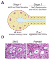
A. Two stage salivary gland secretion model. In Stage 1, acinar cells secrete a NaCl-rich fluid called primary saliva. In Stage 2, the primary saliva is modified through its passage along the duct epithelium by reabsorbing NaCl and secreting KHCO3. Due to the poor permeability of the ducts for water, the resulting final saliva is hypotonic. B. The morphology of human submandibular (left panel) and parotid (right panel) glands. Hematoxylin and Eosin stained submandibular gland section shows that the gland is mainly composed of serous and mucous acinar cells (SA and MA, respectively) and duct cells (Dc). In contrast, human parotid glands are mainly composed of serous acinar cells (SA) as well as duct cells (Dc).
Figure 2
Figure 2
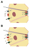
Acinar Cl “pump-leak” models. Fluid secretion by salivary glands is driven by transcellular Cl movement. The primary Cl secretion mechanism relies on the basolateral Na+-K+-2Cl co-transporter (Figure 2A) to mediate Cl influx, whereas the parallel activities of Na+/H+ and Cl/HCO3 exchangers mediate Cl influx in the secondary Cl secretion mechanism (Figure 2B). Primary and secondary Cl uptake (pump) mechanisms depend on the large inward-directed Na+ electrochemical gradient generated by the basolateral Na+/K+ ATPase to promote Na+ extrusion in exchange for extracellular K+, which is recycled into the serosal space by basolateral K+ channels. K+ channel opening also supports Cl secretion (leak) by hyperpolarizing the membrane potential. Cl efflux into the luminal space occurs through apical Cl channels. Apical K+ channels enhance Cl efflux by hyperpolarizing the apical plasma membrane.
Figure 3
Figure 3
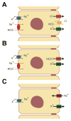
K+ secretion models by salivary gland ducts. The current model for K+ secretion predicts that salivary gland duct cells secrete K+ and HCO3. Two molecular mechanisms have been proposed for K+ - and HCO3 -coupled secretion by salivary gland ducts. A. Primary K+ and HCO3 secretion model, in which basolateral Na+/K+ ATPase promotes K+ accumulation in duct cells above its equilibrium potential (EK) while apical K+ channels are responsible for K+ efflux into the ductal lumen. Na+-HCO3 co-transporters may mediate HCO3 uptake across the basolateral membrane and apical Cl/HCO3 exchangers mediate HCO3 efflux into the ductal lumen. An apical Cl channel recycles Cl ions into the lumen. B. Secondary K+ and HCO3 secretion model is identical to the primary secretion model, except that an apical HCO3 - permeable channel is responsible for the HCO3 efflux into the ductal lumen. Note that apical HCO3 - permeable channel replaces the parallel activities of apical Cl/HCO3 exchangers and Cl channels shown in the primary secretion model. C. In addition to the K+ - and HCO3 -coupled secretion models, there is also experimental evidence supporting a Na+-reabsorptive –dependent K+ secretion process, in which the electrogenic Na+ influx via ENaC channels depolarizes the apical membrane of duct cells thus increasing the driving force for K+ secretion through apical K+ channels. K+ uptake and Na+ efflux across the basolateral membrane occurs via Na+/K+ ATPase.

References

    1. Amerongen AV, Veerman EC. Saliva--the defender of the oral cavity. Oral Dis. 2002;8:12–22. - PubMed
    1. Hay DI, Bowen WH. The functions of salivary proteins. In: Edgar WM, O’Mullane DM, editors. Saliva and oral health. 2. London, UK: British Dental Association; 1996.
    1. Humphrey SP, Williamson RT. A review of saliva: normal composition, flow, and function. J Prosthet Dent. 2001;85:162–169. - PubMed
    1. Mandel ID. Impact of saliva on dental caries. Compend Suppl. 1989:S476–481. - PubMed
    1. Sonies BC, Ship JA, Baum BJ. Relationship between saliva production and oropharyngeal swallow in healthy, different-aged adults. Dysphagia. 1989;4:85–89. - PubMed

Publication types

MeSH terms

Substances