Review
. 2014 Feb 19;81(4):740-54.
doi: 10.1016/j.neuron.2014.01.045.

ApoE and Aβ in Alzheimer's disease: accidental encounters or partners?

Affiliations
Review

ApoE and Aβ in Alzheimer's disease: accidental encounters or partners?

Takahisa Kanekiyo et al. Neuron. .

Abstract

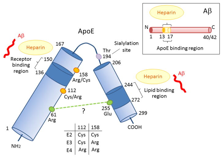
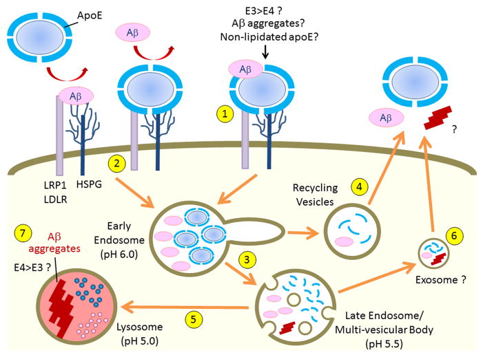
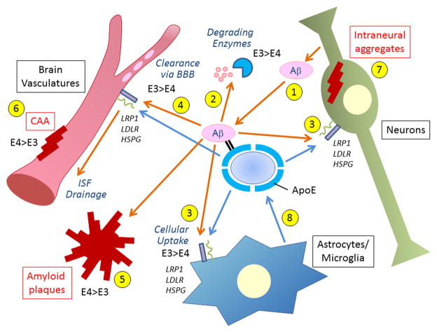
Among the three human apolipoprotein E (apoE) isoforms, apoE4 increases the risk of Alzheimer's disease (AD). While transporting cholesterol is a primary function, apoE also regulates amyloid-β (Aβ) metabolism, aggregation, and deposition. Although earlier work suggests that different affinities of apoE isoforms to Aβ might account for their effects on Aβ clearance, recent studies indicate that apoE also competes with Aβ for cellular uptake through apoE receptors. Thus, several factors probably determine the variable effects apoE has on Aβ. In this Review, we examine biochemical, structural, and functional studies and propose testable models that address the complex mechanisms underlying apoE-Aβ interaction and how apoE4 may increase AD risk and also serve as a target pathway for therapy.


Figures

Figure 1
Figure 1. Schematic illustration of structural and functional regions of apoE and Aβ
Human apoE is a glycosylated protein of 299 amino acids consisting of a receptor-binding region (residues 136–150) in the N-terminal domain (residues 1–167) and a lipid-binding region (residues 244–272) in the C-terminal domain (residues 206–299) (Chen et al., 2011). ApoE also has two heparin-binding sites, each within the N-terminal and the C-terminal domains (Ji et al., 1993; Saito et al., 2003). The residues that distinguish the apoE isoforms are located at residues 112 and 158, where apoE2 has Cys residues at both positions, apoE3 has a Cys residue at 112 and an Arg residue at 158, and apoE4 has Arg residues at both positions (Rall et al., 1982; Weisgraber et al., 1981). The domain interaction between Arg 61 and Glu 255 in apoE4 is also indicated (Mahley and Rall, 2000; Mahley et al., 2009; Wilson et al., 1991), although other structural studies have not confirmed this (Chen et al., 2011). Aβ can interact with both the receptor-binding region and lipid-binding region of apoE, as well as with heparin through its residues 13–17 (Strittmatter et al., 1993; Winkler et al., 1999).
Figure 2
Figure 2. Aβ aggregation: role of apoE
During the Aβ aggregation process, Aβ monomers change their conformation to a β-sheet-rich structure and form soluble oligomers or insoluble intermediate aggregates. Such nuclei further accelerate the fibrillogenesis to form large insoluble fibrils as “seeds” (Harper and Lansbury, 1997). The association of apoE with an Aβ nucleus is likely to block its seeding effect which accelerates Aβ fibrillogenesis. Under certain conditions, apoE and Aβ may form large co-aggregates. Newly generated Aβ fibrils can bind to existing aggregates, resulting in the formation of even larger co-aggregates, with or without additional apoE (Wood et al., 1996a). Finally, these aggregates may deposit as amyloid plaques in the brain.
Figure 3
Figure 3. Cell surface binding and endocytic trafficking of apoE and Aβ
ApoE likely binds to Aβ in an isoform-dependent manner with apoE3 forming more stable apoE/Aβ complexes than apoE4 (LaDu et al., 1994; LaDu et al., 1995). LRP1, LDLR and HSPG are major cell surface receptors that bind apoE, Aβ and apoE/Aβ complexes. In addition to forming a stable complex with Aβ (1), apoE likely competes with Aβ to common cell surface receptors (2) (Verghese et al., 2013). Endocytosed apoE either dissociates from lipid components within the early endosomes due to lower pH (3) and recycles (4), or be transported to lysosomes for degradation (5). Endocytosed Aβ is typically delivered to lysosomes for degradation (5), although a small amount of Aβ can be recycled (4) (Li et al., 2012). In some conditions, apoE and Aβ may be transferred through exosomes from the late endosomes/multi-vesicular body (6). When Aβ accumulation overwhelms the capacity of lysosomes for degradation, the low pH in the lysosomes provide a suitable environment to initiate Aβ aggregation (7) (Hu et al., 2009), which could injure lysosomes and also provide seeding for further Aβ aggregation.
Figure 4
Figure 4. Major Aβ clearance pathways and effects of apoE isoforms
Aβ is predominantly generated in neurons (1) and eliminated through three major clearance pathways including proteolytic degradation by (2) endopeptidases (e.g., NEP, IDE) (Saido and Leissring, 2012), (3) cellular clearance by cells in the brain parenchyma (neurons, astrocytes and microglia) (Kanekiyo et al., 2013; Koistinaho et al., 2004), and (4) ISF drainage where it is degraded by vascular cells (Bell et al., 2009; Kanekiyo et al., 2012) or transported out of the brain through BBB (Ito et al., 2013). Disturbance of these pathways induce Aβ accumulation and deposition in the brain parenchyma as amyloid plaques (5), in the perivascular region as CAA (6) and sometimes also inside neurons (7). ApoE is generated mainly by the glial cells (8) and encounters Aβ in all these pathways. ApoE likely facilitates Aβ clearance by activating enzymatic degradation (2) and phagocytosis (3) in an isoform-dependent manner (apoE3 > apoE4). However, apoE might also suppress Aβ clearance (apoE4 > apoE3) by either competing with Aβ for receptor binding or by retaining Aβ from it clearance through the BBB (4). ApoE4 exacerbates Aβ deposition as amyloid plaques and CAA formation when compared with apoE3 (5 and 6) (Fryer et al., 2005b). LRP1, LDLR and HSPG, which are expressed in all major cellular Aβ clearance pathways, regulate Aβ clearance either directly or through apoE.

Similar articles

Cited by

References

    1. Aguzzi A, Barres BA, Bennett ML. Microglia: scapegoat, saboteur, or something else? Science. 2013;339:156–161. - PMC - PubMed
    1. Baitsch D, Bock HH, Engel T, Telgmann R, Muller-Tidow C, Varga G, Bot M, Herz J, Robenek H, von Eckardstein A, et al. Apolipoprotein E induces antiinflammatory phenotype in macrophages. Arterioscler Thromb Vasc Biol. 2011;31:1160–1168. - PMC - PubMed
    1. Bales KR, Liu F, Wu S, Lin S, Koger D, DeLong C, Hansen JC, Sullivan PM, Paul SM. Human APOE isoform-dependent effects on brain beta-amyloid levels in PDAPP transgenic mice. J Neurosci. 2009;29:6771–6779. - PMC - PubMed
    1. Bales KR, Verina T, Dodel RC, Du Y, Altstiel L, Bender M, Hyslop P, Johnstone EM, Little SP, Cummins DJ, et al. Lack of apolipoprotein E dramatically reduces amyloid beta-peptide deposition. Nat Genet. 1997;17:263–264. - PubMed
    1. Bamberger ME, Harris ME, McDonald DR, Husemann J, Landreth GE. A cell surface receptor complex for fibrillar beta-amyloid mediates microglial activation. J Neurosci. 2003;23:2665–2674. - PMC - PubMed

Publication types