. 2014 Feb 19;81(4):901-12.
doi: 10.1016/j.neuron.2013.12.027.

Dopamine neurons control striatal cholinergic neurons via regionally heterogeneous dopamine and glutamate signaling

Affiliations

Dopamine neurons control striatal cholinergic neurons via regionally heterogeneous dopamine and glutamate signaling

Nao Chuhma et al. Neuron. .

Abstract

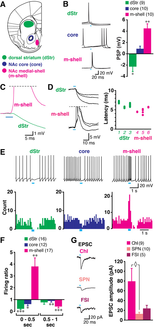
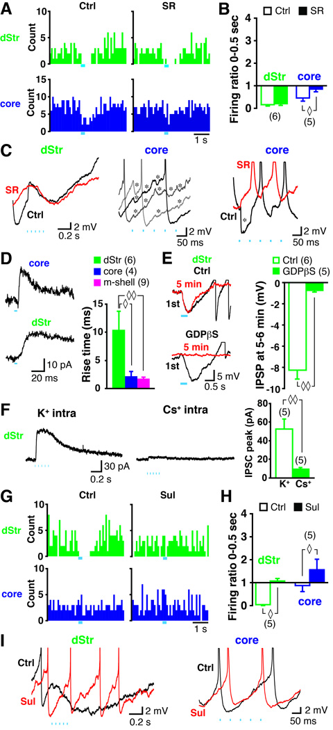
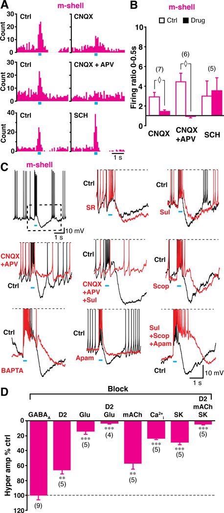
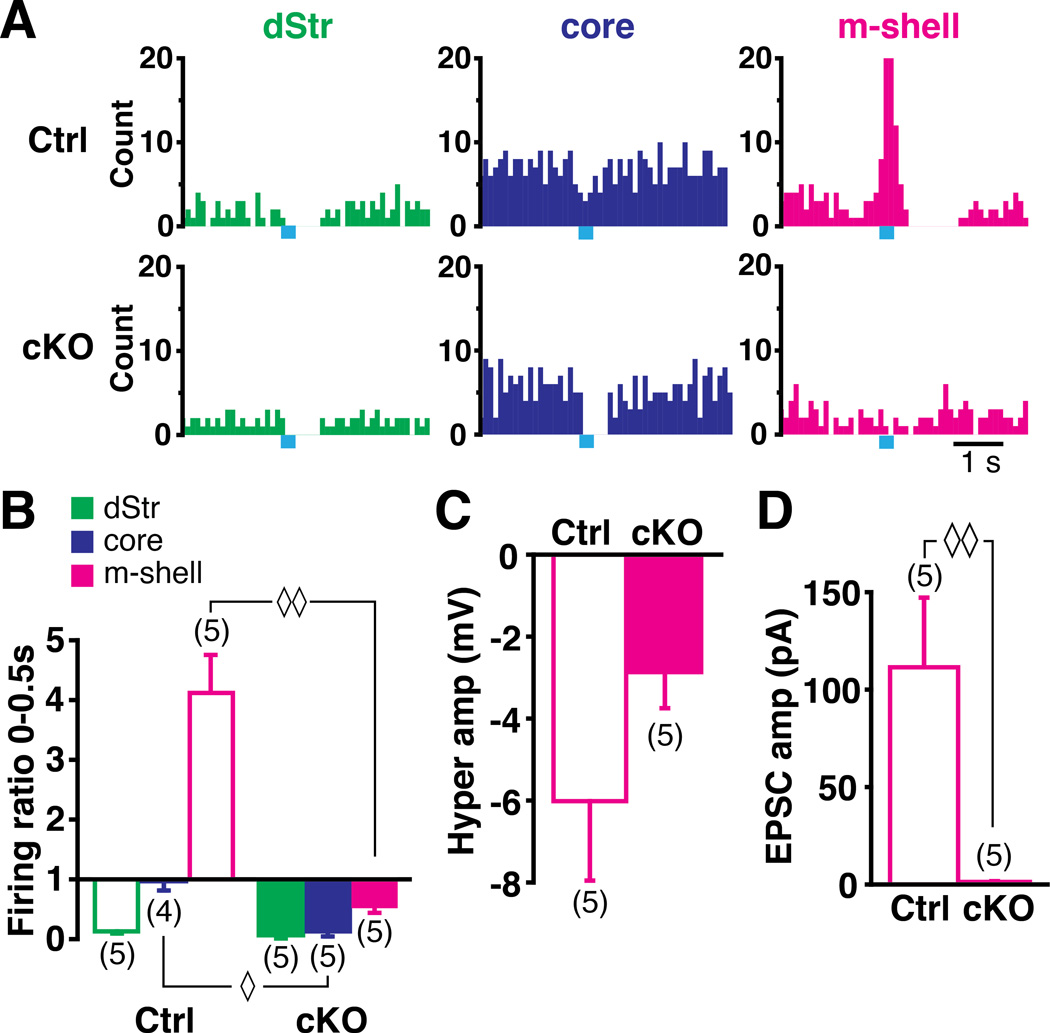
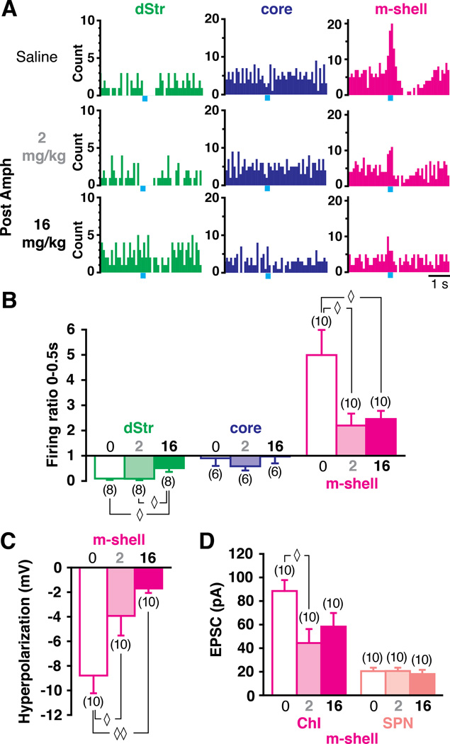
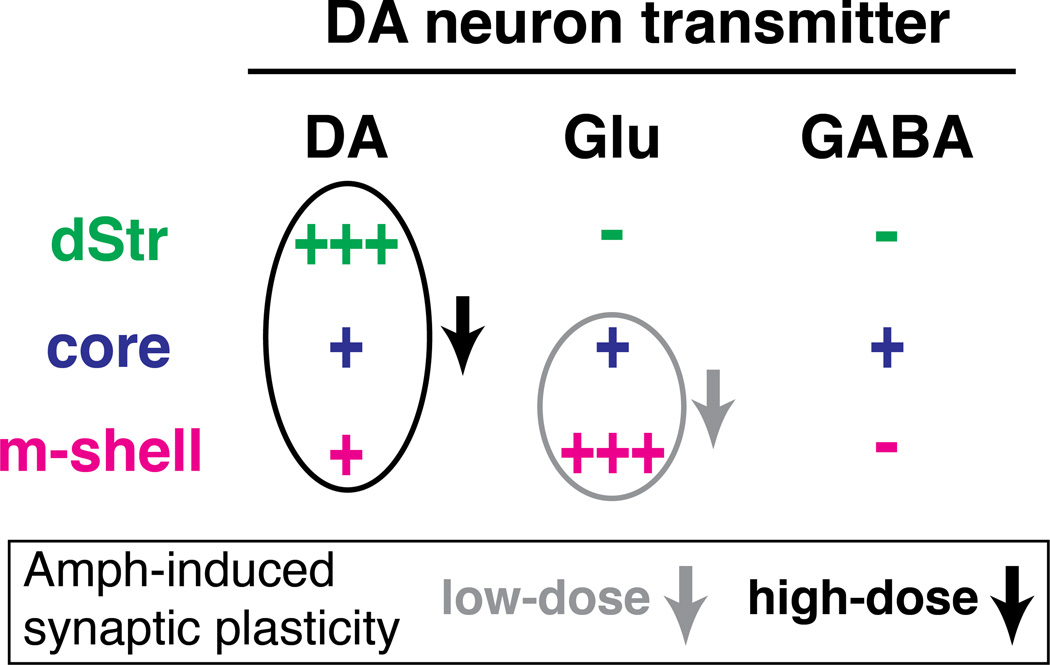
Midbrain dopamine neurons fire in bursts conveying salient information. Bursts are associated with pauses in tonic firing of striatal cholinergic interneurons. Although the reciprocal balance of dopamine and acetylcholine in the striatum is well known, how dopamine neurons control cholinergic neurons has not been elucidated. Here, we show that dopamine neurons make direct fast dopaminergic and glutamatergic connections with cholinergic interneurons, with regional heterogeneity. Dopamine neurons drive a burst-pause firing sequence in cholinergic interneurons in the medial shell of the nucleus accumbens, mixed actions in the accumbens core, and a pause in the dorsal striatum. This heterogeneity is due mainly to regional variation in dopamine-neuron glutamate cotransmission. A single dose of amphetamine attenuates dopamine neuron connections to cholinergic interneurons with dose-dependent regional specificity. Overall, the present data indicate that dopamine neurons control striatal circuit function via discrete, plastic connections with cholinergic interneurons.


Conflict of interest statement

The authors have no conflicts of interest to declare.

Figures

Figure 1
Figure 1. Selective expression of ChR2 in ventral midbrain DA neurons
(A–C) Double immunostaining for YFP (green, left) and TH (magenta, right) of SNc and VTA in AAV-DIO-ChR2-YFP injected DAT-IRES-cre mice (merged images are shown in the middle). (A) The VTA and medial SN are shown at low magnification. (B,C) Representative regions in the SNc (B) and in the VTA (C) are shown at high magnification. Arrows in C indicate a TH positive ChR2 negative neuron. (D) Direct YFP fluorescence is shown in a horizontal slice used for electrophysiological recording. The dashed line indicates the midline. (E) Confirmation of DA neuron responses to train photostimulation, 5 pulses at 20 Hz, in the SN (left) and in the VTA (right). Photostimulation (blue bars) reliably generated action potentials (photostimulation time interval is outlined in dashes and expanded below). See also Fig. S1.
Figure 2
Figure 2. Photostimulation of DA neuron terminals drives regionally heterogeneous synaptic responses in ChIs
(A) Recording were made in the dStr (green), the NAc core (dark blue) and the NAc m-shell (magenta). (B) PSPs evoked by single photostimulation (0.1 Hz, 5 ms duration; blue bars) in ChIs were recorded in the three regions (three traces are shown superimposed); the summary of PSP amplitudes is shown on the right. Negative values indicate hyperpolarization; numbers of recorded cells are in parentheses. PSP amplitudes differing significantly from baseline (0 mV) are indicated respectively by * and ** for p < 0.05 and p < 0.01, by one-sample t-test. (C) Expansion of the onset of PSPs recorded in the dStr and the m-shell is shown. Averages of 10 traces are shown. Action potentials are truncated (horizontal dashed line). (D) Onset of three individual PSPs is shown superimposed, with the latency from the onset of photostimulation summarized for 3 cells in each region. Cell identification number (1–6) is in italics. Each closed circle is the measurement from a single trace; 8–10 traces were measured per cell (many circles overlap). (E) Effect on ChI firing mimicking DA neuron bursting (5 pulses at 20 Hz) in the three regions. Sample traces from each region are shown (top), with peristimulus histograms made from 10 consecutive traces (0.1 sec bin) (below). Blue bars indicate timing of train photostimulation. (F) Ratio of firing during train (0–0.5 sec from onset of train, left) and just after train (0.5–1 sec from onset, right) to baseline firing (firing ratio). Significant changes from baseline firing (ratio 1) are indicated by *, ** and *** for p < 0.05, p < 0.01 and p < 0.001 by one-sample t-test, respectively. (G) Comparison of fast EPSCs generated by single pulse photostimulation among three cell types in the m-shell. Sample traces recorded from a ChI, SPN and FSI are shown (left); traces are averages of 10 consecutive traces. In a summary of EPSC amplitudes (right), ◊ indicates p < 0.05 by one-way ANOVA with Scheffe’s post-hoc test. Data are presented as mean ± s.e.m. See also Fig. S2.
Figure 3
Figure 3. Neurotransmitter mediation of the pause in the dStr and firing attenuation in the core
(A–C) Effect of the GABAA antagonist SR95531 (10 µM). Peristimulus histograms of firing under control (Ctrl) and after application of SR95531 (SR), in the dStr and the core are shown (A). Blue bars indicate train photostimulation. In a summary of firing ratios (B), ◊ indicates p < 0.05 by Wilcoxon signed ranks test. Numbers of recorded cells are in parentheses. Expanded sample traces (C) of responses to train photostimulation are shown for control (black) and after application of SR95531 (red), in the dStr and in the core. Middle panel shows three superimposed control traces with probable GABAergic IPSPs (*). Action potentials were truncated. (D–F) The hyperpolarization in the dStr was G-protein coupled and K+ channel mediated. Sample traces of IPSCs evoked with a single light pulse are shown for the core and dStr (D, left). IPSCs are the average of 10 traces. In measurements of the rise time (from 10 – 90% peak amplitude; D, right), ◊ and ◊◊ indicate p < 0.05 and p < 0.01, respectively, by one-way ANOVA with Scheffe’s posthoc test. Intracellular 1 –1.5 mM GDPpS blocked the IPSP in the dStr (E). The first trace (black) and the trace 5 min after entering whole cell mode (red) are shown with control intracellular solution and with GDPpS (E, left). In a comparison of average IPSP amplitude at 5 – 6 min in control and with GDPpS (E, right), ◊◊ indicates p < 0.01 by Mann-Whitney U test. With Cs+-based intracellular solution, IPSCs (holding at −60 mV) were blocked (F). IPSC traces at 5 min after entering whole cell mode evoked by a train photostimulation with K+-(F, left) and Cs+-(F, middle) intracellular solution are shown. In a comparison of IPSC peak amplitude at 5 – 6 min after entering whole cell mode (F, right), ◊◊ indicates p < 0.01 by Mann-Whitney U test. (G–I) Effect of D2 antagonist sulpiride (10 µM). Peristimulus histograms of firing under control and after application of sulpiride, in the dStr and the core are shown (G). In a summary of firing ratios (H), ◊ indicates p < 0.05 by Wilcoxon signed ranks test. Expanded sample traces for control (black) and after sulpiride (red) are shown. See also Fig. S3.
Figure 4
Figure 4. Neurotransmitter mediation of burst and PBH in the m-shell
(A–B) Pharmacology of burst. Effects of AMPA/kainate antagonist CNQX (40 µM), a cocktail of CNQX + NMDA antagonist D-APV (100 µM) and D1 antagonist SCH23390 (10 µM) are shown in peristimulus histograms (A) and firing ratios (B). ◊ indicates p < 0.05 by Wilcoxon signed ranks test. (C–D) Pharmacology of PBH. The first sample trace shows the burst followed by PBH (C, top left). The PBH (dashed rectangle outline) is expanded in the subsequent panels. Sample traces show control PBH (black) and after drug application (red). Drugs tested were (from top middle to bottom right) SR95531, sulpiride, a cocktail of CNQX + D-APV, CNQX+D-APV+sulpiride, scopolamine, intracellular BAPTA, apamin, and a cocktail of sulpiride+scopolamine+apamin. The percent reduction in the PBH amplitude for each of the drugs is shown (D). The dashed line indicates control (no change). ** and *** indicate p < 0.01 and p < 0.001, respectively, with one-sample t-test.
Figure 5
Figure 5. cKO of VGLUT2 in DA neurons eliminates burst firing and reduces regional heterogeneity
(A) Peristimulus histograms of firing in the three regions in control (top) and in VGLUT2 cKO (bottom) slices. (B) Firing ratio comparison between control and cKO in three regions. (C) PBH amplitude appears diminished in cKO, but this was not significant. (D) EPSCs evoked by single pulse photostimulation are absent in cKO, showing the complete block of DA neuron glutamate cotransmission in cKO. ◊ and ◊◊ indicate p < 0.05 and p < 0.01, respectively, by Mann-Whitney U test.
Figure 6
Figure 6. Amphetamine-induced plasticity is regionally heterogeneous
(A) Peristimulus histograms of firing in the three regions in mice previously treated with saline (top), low-dose (middle) and high-dose (bottom) amphetamine. (B) Firing ratio measurements show that following low-dose ChI bursts are attenuated selectively in the m-shell. (C) The PBH in the m-shell shows a progressive dose-dependent reduction. (D) EPSCs evoked by single-pulse photostimulation (at 0.1 Hz) in m-shell ChIs were attenuated following amphetamine, while there was no effect in SPNs. ◊ and ◊◊ indicate p < 0.05 and p < 0.01, respectively, by one-way ANOVA with Scheffe’s post-hoc test. See also Fig. S4.
Figure 7
Figure 7. Regional heterogeneity of fast DA neuron transmission to ChIs is accentuated by amphetamine
Fast synaptic actions of DA neurons on ChIs are mediated by DA, glutamate and GABA. The strength of connections is indicated by the number of plus signs. Ovals and arrows indicate differential attenuation of synaptic effects following low-dose or high-dose amphetamine.

References

    1. Araya R, Andino-Pavlovsky V, Yuste R, Etchenique R. Two-photon optical interrogation of individual dendritic spines with caged dopamine. ACS Chem Neurosci. 2013;4:1163–1167. - PMC - PubMed
    1. Atasoy D, Aponte Y, Su HH, Sternson SM. A FLEX switch targets Channelrhodopsin-2 to multiple cell types for imaging and long-range circuit mapping. J Neurosci. 2008;28:7025–7030. - PMC - PubMed
    1. Bäckman CM, Malik N, Zhang Y, Shan L, Grinberg A, Hoffer BJ, Westphal H, Tomac AC. Characterization of a mouse strain expressing Cre recombinase from the 3' untranslated region of the dopamine transporter locus. Genesis. 2006;44:383–390. - PubMed
    1. Beckstead MJ, Grandy DK, Wickman K, Williams JT. Vesicular dopamine release elicits an inhibitory postsynaptic current in midbrain dopamine neurons. Neuron. 2004;42:939–946. - PubMed
    1. Belin D, Jonkman S, Dickinson A, Robbins TW, Everitt BJ. Parallel and interactive learning processes within the basal ganglia: relevance for the understanding of addiction. Behav Brain Res. 2009;199:89–102. - PubMed

Publication types

LinkOut - more resources