Review
. 2014:123:53-82.
doi: 10.1016/B978-0-12-397897-4.00011-5.

Implications of cellular models of dopamine neurons for schizophrenia

Affiliations
Review

Implications of cellular models of dopamine neurons for schizophrenia

Na Yu et al. Prog Mol Biol Transl Sci. 2014.

Abstract

Midbrain dopamine neurons are pacemakers in vitro, but in vivo they fire less regularly and occasionally in bursts that can lead to a temporary cessation in firing produced by depolarization block. The therapeutic efficacy of antipsychotic drugs used to treat the positive symptoms of schizophrenia has been attributed to their ability to induce depolarization block within a subpopulation of dopamine neurons. We summarize the results of experiments characterizing the physiological mechanisms underlying the ability of these neurons to enter depolarization block in vitro, and our computational simulations of those experiments. We suggest that the inactivation of voltage-dependent Na(+) channels, and, in particular, the slower component of this inactivation, is critical in controlling entry into depolarization block. In addition, an ether-a-go-related gene (ERG) K(+) current also appears to be involved by delaying entry into and speeding recovery from depolarization block. Since many antipsychotic drugs share the ability to block this current, ERG channels may contribute to the therapeutic effects of these drugs.

Keywords: Antipsychotic drugs; Bursting; Depolarization block; Ether-a-go-go-related gene (ERG) potassium current; K(v)11; Pacemaker; Substantia nigra; Ventral tegmental area.


Figures

Figure 1
Figure 1
Intracellular recordings of spontaneously firing DA neurons in haloperidol-treated rats. Although most of the DA neurons in treated rats are not spontaneously active, a few firing neurons may nonetheless be impaled with a recording electrode. These neurons typically demonstrate rapid, high-amplitude burst activity unlike that commonly observed in DA neurons in control animals. A, rapid, high-amplitude bursting in a DA cell presumably on the threshold of depolarization block. B, a DA cell in a less depolarized state. Final spikes in burst are inactivated. Reproduced with permission from Figure 2 of Grace and Bunney 1986 with the original figure caption.
Figure 2
Figure 2
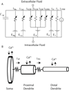
Model of dopamine neuron as a spiking pacemaker. A. Conductance-based equivalent circuit for each compartment (Istim is confined to the soma). The arrows indicate time and voltage dependent conductances. B. Schematic representation of the multi-compartmental model and the calcium balance.
Figure 3
Figure 3
Currents contributing to AHP and ISI duration. A and B show the somatic membrane potential during spontaneous spiking activity, simulated from schematic DA neuron model. Also indicated is the portion of the interspike interval (ISI) analyzed below. C and D show the most important currents that contribute to the total somatic current during the time intervals of the ISI indicated in panels A and B. Labeling: IKA, hyperpolarization activated K+ current; INaP, electrogenic Na+ pump; ICa,L, Ca2+ L-type channel current, ISP dendritic current; IL, leak current; ISoma, net contribution from IKA, INaP, ICa,L, ISP and IL to total somatic current is indicated by solid curve. The total somatic current was obtained by multiplying the somatic current densities in the model by the somatic surface area. Reproduced from Figure 3 of Kuznetsova et al., 2010 with kind permission from Springer Science and Business Media.
Figure 4
Figure 4
Simulated induction of depolarization block by simulated somatic current injection. A: Somatic membrane potential trace from schematic DA neuron model under control conditions before simulated applied current and during application of 500 pA pulse. In the latter case a cessation of the spiking response is observed. B) Inactivation variable (h) of fast Na+ current as a function of time for the simulation in A. At the larger value of applied current the inactivation of Na+ current is not removed leading to depolarization block. Adapted with permission from Figure 6 in Kuznetsova et al., 2010.
Figure 5
Figure 5
Spiking current conductance densities control the maximum frequency in the schematic model. Frequency current plots at different levels of sodium conductance density (gNa). Adapted with permission from Figure 8 (a) in Kuznetsova et al., 2010.
Figure 6
Figure 6
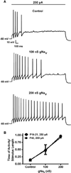
Additional somatic NaV channels reduce DP block produced by current injection in adolescent and older animals. Representative recordings from SNc DA neurons current-clamped at −60 mV followed by (A) a 2 s, 250 pA current injection to induce DP block under control conditions (top) and with 100 nS (middle) or 200 nS (bottom) of virtual NaV added via dynamic clamp. B) Resistance to DP block as quantified by length of activity divided by the stimulus length plotted against the virtual NaV conductance in response to a current injection. Current injections were 250 pA for SN DA neurons in slices from P14 –P21 rats (open circles; n=11) and 200 pA current injection to SN DA neuron in slices from a P42 rat (black square; n=3). Symbols and error bars indicate the mean ± SEM. Adapted with permission from Figure 5 Tucker et al., 2012.
Figure 7
Figure 7
Voltage-dependent characteristics of ERG K+ channels. A: Cartoon schematic illustrating the three conductance states of ERG K+ channels. B: Macroscopic current corresponding to the conductance states in illustrated A. At hyperpolarized membrane potentials, ERG channels are closed (C). Depolarizing the membrane potential from −80 mV to 0 mV (lower trace) slowly opens the channels (C → O) resulting in an initial outward current as K+ ions diffuse out of the cell. However, ERG channels rapidly inactivate (O → I) by entering a second non-conductive state that limits the outward current produced by the initial depolarization. Partial repolarization of the membrane potential induces a rapid transition from an inactive to an open conformation (I → O). Since the rate of deinactivation (I →O) exceeds the rate at which the channels can close, a large ‘resurgent’ current is generated as the membrane potential repolarizes. Reprinted with permission from Figure 1 of Shepard et al., 2007.
Figure 8
Figure 8
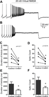
The prototypical ERG channel blocker E-4031 promotes depolarization block by virtual NMDA receptor stimulation. A and B are representative whole-cell dynamic-clamp recordings of DA neurons stimulated with 20 nS of virtual NMDA current for 5 seconds to induce depolarization block before (A) and after (B) superfusion with 10 µM E-4031. The horizontal line above the traces indicates virtual NMDA conductance application. Summary of the effects of 10 µM E-4031 on the duration (C,E) and spike count (D,F) from dynamic clamp simulations of NMDA-induced bursting activity. *p<0.05. Adapted with permission from Figure 4 of Ji et al., 2012.
Figure 9
Figure 9
ERG K+ channel block prolongs plateau potentials induced by the SK channel negative modulator, NS8593. A: Representative tracing from a DA neuron in the presence of NS8593 (6 uM). Note the “burst” of action potentials elicited during the initial phase of a depolarizing plateau potential. B: Addition of E-4031 (10 µM) prolongs the duration of the plateau. C: Box (25th and 74th percentiles) and whisker (10th and 90th percentiles) plot illustrating the effects of E-4031 (10 µM) on the duration of spontaneous plateau potentials elicited by negative modulation of SK channels. Solid and dashed lines inside the box represent the median and mean, respectively. *** P < 0.003 vs. NS8593. D: Perfusion with E-4031 (10 µM) results in the loss of spontaneous activity through depolarization block following removal of a negative bias current. Brief hyperpolarizing current pulses (0.03 nA, 200 ms; vertical arrows) are capable of repolarizing the neuron which leads to recovery of spontaneous spiking followed by the rapid return of depolarization block. Adapted with permission from Figure 3 of Ji et al., 2012.
Figure 10
Figure 10
Model of dopamine neuron extended to simulate bursting and pacing. Details are given in the appendix.
Figure 11
Figure 11
How additional NaV conductance rescues depolarization block. A1) Steady state voltage dependence for fast inactivation (h) and slow inactivation (hs). A2) The peak of the sodium current elicited by a10 Hz train of 3 msec voltage steps from −70 mV to 0 mV decays during the train due to accumulation of slow inactivation hs. This simulation mimics the protocol of Figure 8 in Ding et al., 2011. B) This simulation mimics the protocol in Figure 6 in which a 2 second current pulse is applied. B1) Under control conditions, the pulse causes entry into depolarization block. B2) The simulated addition of virtual NaV conductance prevents entry into depolarization block. B3) The additional conductance allows for more recovery from slow inactivation. B4) At similar levels of hs, the recovery under control from h is similar to that with added NaV, but deteriorates as the difference in hs is established. B5) The major difference is that the noninactivated (available) conductance after each spike is increased with additional conductance.
Figure 12
Figure 12
How the ERG current delays entry into depolarization block. A, Top) A square pulse of simulated virtual NMDA conductance applied to a pacemaking model neuron induces a burst of spikes followed by depolarization block. A, Bottom) Simulated ERG current block increases the pacemaker rate, and accelerates entry into depolarization block, shortening both the bursting duration and the number of spikes in the burst. B) ERG block clearly decreases recovery from slow sodium channel inactivation (hs) during the square pulse in conductance with a more ambiguous effect on fast sodium inactivation (h). The addition of ERG channels enhances hs, thus the fraction of available sodium channels (h*hs). C) During a single spike, the potential during the interspike interval is elevated in the absence of the ERG current (dashed trace) compared to control (solid trace), resulting in more slow inactivation (lower levels of hs). The change in the level of h due to a spike is greater under control conditions due to the extra after hyperpolarization due to the ERG current. The time course of the ERG current and its open state (o), inactivation state (i) is shown for the control case only.
Figure 13
Figure 13
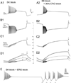
How the ERG current contributes to plateau repolarization. A1) Simulated block of the SK current produces a burst of spikes followed by DP block. A2) A 30% reduction in the ERG conductance again shortens the burst of spikes and also prolongs the depolarized plateau. B) The time course of the L-type Ca2+ current, with the arrow indicating the regenerative decrease in this current that drives the sharp plateau repolarization in A. C) The time course of the ERG current, with the arrow showing the point at which this current tips the balance in favor of the outward currents to initiate plateau repolarization. D) The ERG channel fraction in the open state (o) and inactivation state (i) as well as the slow pool of o+i (bold gray curve) show the slow time course of the combined pool as well as the bump in the open state during repolarization. E) A complete block of the ERG channels induces permanent depolarization block at −40mV. Brief (200 ms) hyperpolarizing somatic current pulses (gray bars) temporarily relieve depolarization block

References

    1. Bernheimer H, Birkmayer W, Hornykiewicz O, Jellinger K, Seitelberger F. Brain dopamine and the syndromes of Parkinson and Huntingdon: Clinical, morphological, and neurochemical correlations. J Neurosci. 1973;20:415–455. - PubMed
    1. Bloom FE, Roth RH, Cooper JR. The biochemical basis of neuropharmacology. Oxford: Oxford University Press; 1991.
    1. Bunney BS, Grace AA. Acute and chronic haloperidol treatment: comparison of effects on nigral dopaminergic cell activity. Life Sci. 1978;23:1715–1727. - PubMed
    1. Canavier CC, Oprisan SA, Callaway J, Ji H, Shepard PD. Computational model predicts a role for ERG current in repolarizing plateau potentials in dopamine neurons: implications for modulation of neuronal activity. J. Neurophysiol. 2007;98:3006–3022. - PubMed
    1. Carlsson A, Lindqvist M. Effect of chlorpromazine or haloperidol on formation of 3-methoxytyramine and normetanephrine in mouse brain. Acta Pharmacol. Toxicol. (Copenh) 1963;20:140–144. - PubMed

Publication types

MeSH terms

Substances