. 2014 Mar 3;24(5):484-93.
doi: 10.1016/j.cub.2014.01.022. Epub 2014 Feb 20.

Evoked and spontaneous transmission favored by distinct sets of synapses

Affiliations

Evoked and spontaneous transmission favored by distinct sets of synapses

Einat S Peled et al. Curr Biol. .

Abstract

Background: Spontaneous "miniature" transmitter release takes place at low rates at all synapses. Long thought of as an unavoidable leak, spontaneous release has recently been suggested to be mediated by distinct pre- and postsynaptic molecular machineries and to have a specialized role in setting up and adjusting neuronal circuits. It remains unclear how spontaneous and evoked transmission are related at individual synapses, how they are distributed spatially when an axon makes multiple contacts with a target, and whether they are commonly regulated.

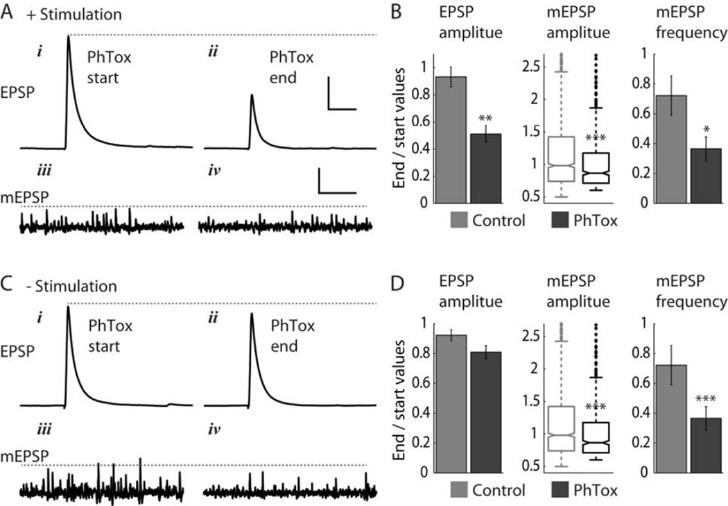
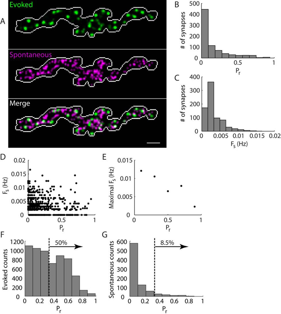
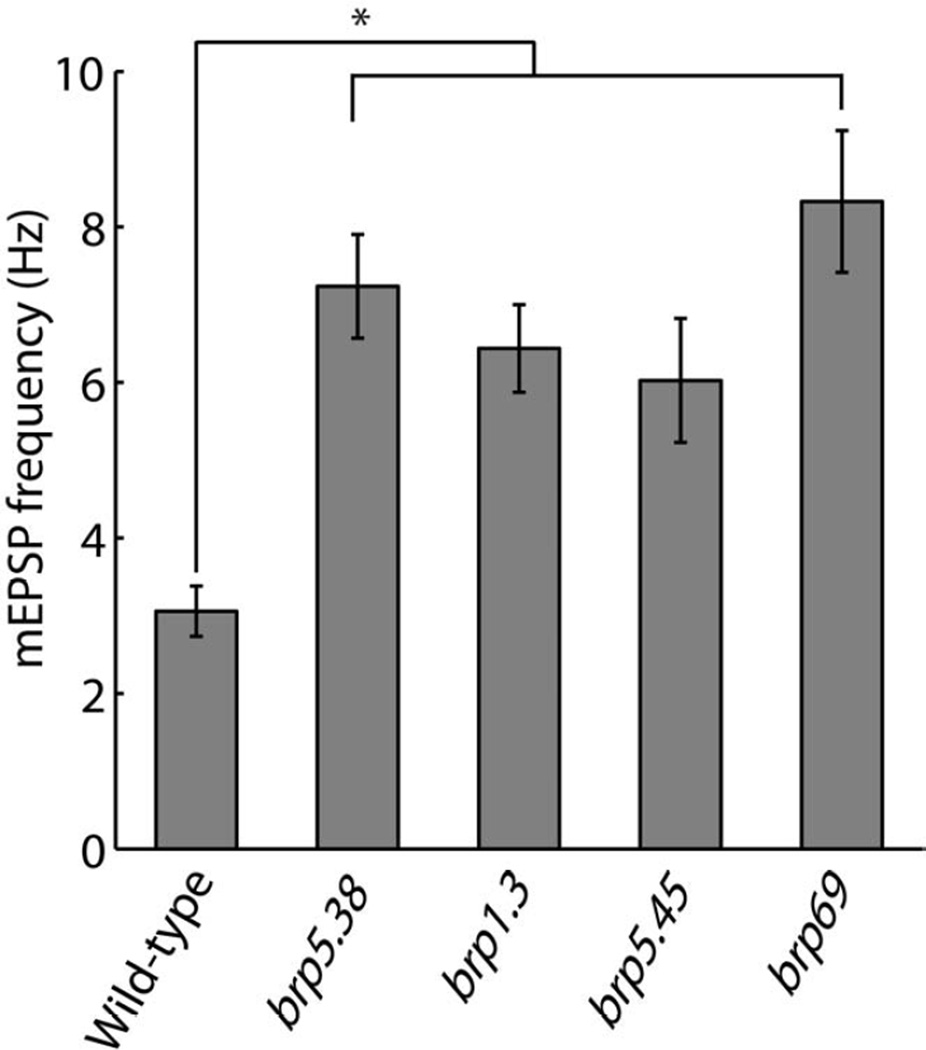
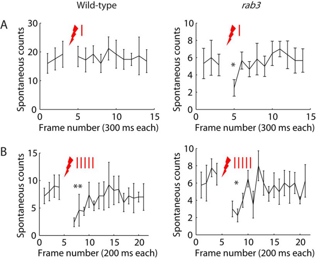
Results: Electrophysiological recordings in the Drosophila larval neuromuscular junction, in the presence of the use-dependent glutamate receptor (GluR) blocker philanthotoxin, indicated that spontaneous and evoked transmission employ distinct sets of GluRs. In vivo imaging of transmission using synaptically targeted GCaMP3 to detect Ca(2+) influx through the GluRs revealed little spatial overlap between synapses participating in spontaneous and evoked transmission. Spontaneous and evoked transmission were oppositely correlated with presynaptic levels of the protein Brp: synapses with high Brp favored evoked transmission, whereas synapses with low Brp were more active spontaneously. High-frequency stimulation did not increase the overlap between evoked and spontaneous transmission, and instead decreased the rate of spontaneous release from synapses that were highly active in evoked transmission.

Conclusions: Although individual synapses can participate in both evoked and spontaneous transmission, highly active synapses show a preference for one mode of transmission. The presynaptic protein Brp promotes evoked transmission and suppresses spontaneous release. These findings suggest the existence of presynaptic mechanisms that promote synaptic specialization to either evoked or spontaneous transmission.


Figures

Figure 1
Figure 1. PhTox blocks spontaneous mEPSPs independently of evoked EPSPs
(A, B) PhTox block during 0.1 Hz nerve stimulation. (Aiiv) Representative experiment showing EPSP and mEPSPs traces at start of PhTox application and 25 minutes later. (B) Quantification of block of EPSP amplitude, mEPSP amplitude, and mEPSP frequency. PhTox n = 5 NMJs, control n = 5 NMJs. (C, D) PhTox block without nerve stimulation (brief 0.1 Hz stimulation was applied only at start and end of experiments). Panels are as in (A, B). PhTox n = 8 NMJs, control n = 7 NMJs. Scale bars in (A, C): 100 ms and 10 mV (EPSP traces), 2 s and 0.5mV (mEPSP traces). Values reported in (B, D) are mean ± SEM (EPSP amplitude and mESPS frequency) and median and first and third quartiles (mEPSP amplitude). Statistical significance tests are Student’s t-test (EPSP amplitude and mEPSP frequency) and Wilcoxon rank sum test (mEPSP amplitude). Asterisks mark significance values: * p < 0.05, ** p < 0.003, *** p < 0.0002. See also Figure S1.
Figure 2
Figure 2. Contrast between evoked and spontaneous transmission in wild-type NMJs
(A) Activity maps showing the probability of synapses to participate in evoked transmission (green) and the rate of spontaneous miniature transmission (magenta) across a wild-type NMJ. Overlap between the two patterns of activity shows in white color in the merged activity map. Maximal observed values for the NMJ shown were Pr = 0.31 and Fs = 0.023 Hz. Correlation coefficient between the activity patterns was 0.32. (B) Blowup of activity maps for the two boutons boxed in (A). Yellow arrows point to locations that show overlap between evoked and spontaneous activity patterns. (C, D) Histograms of Pr (C) and Fs (D) values in wild-type NMJs. (E) Plot of Fs versus Pr for all synapses examined. (F) Plot of maximal observed Fs over three Pr ranges. Maximal values were obtained by dividing Pr to bins of size 0.2 and calculating the mean of the top 10 Fs values for each bin. (G, H) Histograms showing counts of evoked (G) and spontaneous (H) transmission events versus Pr. Dashed line marks the median Pr for evoked counts, Pr = 0.07. 50% of evoked and 20% of spontaneous miniature transmission events occurred at synapses with Pr > 0.07. (C – H) Histograms show data pooled from 957 synapses at 5 wild-type NMJs. Scale bar in (A), 5 μm. See also Figures S2, S3, S4, and Movie S1.
Figure 3
Figure 3. Contrast between evoked and spontaneous transmission in rab3-mutant NMJs
(A) Activity maps showing the probability of synapses to participate in evoked transmission (green) and the rate of spontaneous miniature transmission (magenta) across a rab3-muatnt NMJ. Overlap between the two patterns of activity shows in white color in the merged activity map. Maximal observed values for the NMJ shown were Pr = 0.9 and Fs = 0.017 Hz. Correlation coefficient between the activity patterns was 0.19. (B, C) Histograms of Pr (B) and Fs (C) values in rab3-mutant NMJs. (D) Plot of Fs versus Pr for all synapses examined. (E) Plot of maximal observed Fs over five Pr ranges. Maximal values were obtained by dividing Pr to bins of size 0.2 and calculating the mean of the top 10 Fs values for each bin. (F, G) Histograms showing counts of evoked (F) and spontaneous (G) transmission events versus Pr. Dashed line marks the median Pr for evoked counts, Pr = 0.33. 50% of evoked and 8.5% of spontaneous miniature transmission events occurred at synapses with Pr > 0.33. (B – G) include data pooled from 785 synapses at 6 rab3-NMJs. Scale bar in (A), 5 μm.
Figure 4
Figure 4. Evoked and spontaneous transmission are oppositely correlated with levels of the presynaptic protein Brp
(A) Comparison of Brp staining to locations of evoked and spontaneous transmission in the NMJ. Locations where transmission was detected during live imaging are marked in white on top of a Brp staining image. Note that each marked location typically experienced multiple transmission events over the time course of the experiment. (B) Correlation of single-synapse presynaptic Brp levels with Pr (left) and Fs (right). Reported values are mean ± SEM. Figures include data from 729 synapses (showing either evoked or spontaneous transmission, or presynaptic Brp with no transmission) at 6 rab3-mutant NMJs (Student’s t-test, ** p < 0.006). Scale bar in (A), 5 µm. See also Figure S5.
Figure 5
Figure 5. Increased mEPSP frequency in brp mutants
mEPSP frequency in wild-type and brp-mutant NMJs. All brp mutants had one copy of the indicated mutant allele over the corresponding second-chromosome deficiency BSC29. All brp mutants show elevated mEPSP frequency significantly higher than wild-type values. Reported values are mean ± SEM. n = 16, 12, 10, 10 and 8 for wild-type, brp5.38, brp1.3, brp5.45 and brp69 NMJs, respectively. One-way ANOVA p < 1.1×10⁻⁷. Asterisk marks pairwise significance from post-hoc comparison with Bonferroni criterion (* p < 0.01). See also Figure S6 and Movies S2 and S3.
Figure 6
Figure 6. High-frequency stimulation does not alter overlap between evoked and spontaneous transmission
(A) Merged activity map showing the probability that synapses distributed throughout a wild-type NMJ participate in evoked transmission triggered either by one nerve stimulation (green), five stimulations (magenta), or both (white). Maximal observed values for this NMJ were Pr(1) = 0.7 and Pr(5) = 0.94. The correlation coefficient between these activity patterns was 0.84. (B) Plot of Pr(5) versus Pr(1) for all synapses in the NMJ of (A). Solid line indicates no change in probability (i.e. Pr(5) = Pr(1)). (C) Merged activity maps for the NMJ of (A), showing the probability that synapses participate in either evoked (green) or spontaneous (magenta) transmission or both (white). Right and left panels correspond to one and five nerve stimulations, respectively. For this NMJ, correlation values between the evoked and spontaneous activity maps were 0.36 and 0.24 for one and five nerve stimulations, respectively. Scale bar in (A), 5 µm. See also Figure S7.
Figure 7
Figure 7. Counts of spontaneous miniature events before and after nerve stimulation
Plots showing the number of spontaneous miniature events identified at each SynapGCaMP3 imaging frame versus frame number. Red lightning bolts mark time of nerve stimulation. (A) Single nerve stimulation. Wild-type (left, n = 5) and rab3-mutant (right, n = 6) NMJs. Time between start of consecutive frames was 300 ms. The single nerve stimulation was applied after frame number 3. (B) Five nerve stimulations at 20 Hz. Wild-type (left, n = 5) and rab3-mutant (right, n = 4) NMJs. Time between start of consecutive frames was 200 ms. The train of five nerve stimulations was applied after frame number 4. Reported values are mean ± SEM. Student’s paired t-tests were used to compare spontaneous counts before and after nerve stimulation: (A) comparing one frame (~ 300 ms) before to one frame after nerve stimulation, (B) comparing three frames (~600 ms) before to three frames after nerve stimulation. Asterisks mark significance values: * p < 0.05, ** p < 0.002.

Comment in

Similar articles

Cited by

References

    1. Fatt P, Katz B. Spontaneous subthreshold activity at motor nerve endings. J Physiol. 1952;117:109–128. - PMC - PubMed
    1. Del Castillo J, Katz B. Quantal components of the endplate potential. J Physiol. 1954;124:560–573. - PMC - PubMed
    1. Zucker RS. Minis: whence and wherefore? Neuron. 2005;45:482–484. - PubMed
    1. Kavalali ET, Chung C, Khvotchev M, Leitz J, Nosyreva E, Raingo J, Ramirez DM. Spontaneous neurotransmission: an independent pathway for neuronal signaling? Physiology (Bethesda) 2011;26:45–53. - PubMed
    1. Ramirez DM, Kavalali ET. Differential regulation of spontaneous and evoked neurotransmitter release at central synapses. Curr Opin Neurobiol. 2011;21:275–282. - PMC - PubMed

Publication types

MeSH terms