Review
. 2014 Jun;60(6):1290-303.
doi: 10.1016/j.jhep.2014.02.006. Epub 2014 Feb 19.

Characterization of animal models for primary sclerosing cholangitis (PSC)

Affiliations
Review

Characterization of animal models for primary sclerosing cholangitis (PSC)

Peter Fickert et al. J Hepatol. 2014 Jun.

Abstract

Primary sclerosing cholangitis (PSC) is a chronic cholangiopathy characterized by biliary fibrosis, development of cholestasis and end stage liver disease, high risk of malignancy, and frequent need for liver transplantation. The poor understanding of its pathogenesis is also reflected in the lack of effective medical treatment. Well-characterized animal models are utterly needed to develop novel pathogenetic concepts and study new treatment strategies. Currently there is no consensus on how to evaluate and characterize potential PSC models, which makes direct comparison of experimental results and effective exchange of study material between research groups difficult. The International Primary Sclerosing Cholangitis Study Group (IPSCSG) has therefore summarized these key issues in a position paper proposing standard requirements for the study of animal models of PSC.

Keywords: Animal model; Bile acids; Biliary fibrosis; Cholangiopathies; Cholestatic liver disease; Primary sclerosing cholangitis.


Conflict of interest statement

Conflict of interest: The authors who have taken part in this study declared that they do not have anything to disclose regarding funding or conflict of interest with respect to this manuscript.

Figures

Fig. 1
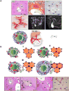
Fig. 1. PSC characteristics and attributes of an ideal PSC animal model
(A) Attributes of an ideal PSC animal model. Ideally onionskin-type periductal fibrosis of intrahepatic bile ducts, bile duct replacement by a scar formation (as illustrated by the asterisk on SR stained section) and development of bile duct proliferation (visualized with K19 staining) as shown in human PSC in the upper panel would be mirrored in such a model. Typical macroscopic appearance of the biliary tree with strictures and dilatations of large and medium-sized bile ducts shown with bile duct plastination of an Abcb4−/− mouse (left) as well as via MRCP in a PSC patient (central panel). In addition, special immunological phenotypes of inflammatory cells infiltrating portal tracts (similar to the human situation) as well as atrophy of cholangiocytes should be present as illustrated by the cartoon (lower panel, left). An association with inflammatory bowel disease together with male predominance (lower panel) would round off the ideal PSC model. Original magnification for the upper panel 100× and 200×. bd, bile duct; cv, central vein; pv, portal vein. (B) Histological changes in human PSC. The characteristics of early stage (stage I and II) include a diffuse mixed cell inflammatory infiltrate around the bile ducts, portal edema, ductular reaction and invading neutrophilic granulocytes (biliary interphase activity) as illustrated by the cartoons (upper panel) and by the HE- and SR-stained sections of human PSC livers (lower panel). Further progression of the disease is accompanied by increasing portal fibrosis, ductular reaction (indicated by the arrows), bile duct replacement by a scar formation (as illustrated by the asterisks) with the formation of portal-portal linking septa (biliary fibrosis) (stage III) and finally the development of cirrhosis (stage IV). Original magnification for the lower panel 100× and 40×. bd, bile duct; cv, central vein; dr, ductular reaction; pv, portal vein.
Fig. 2
Fig. 2. Liver preparation and processing
Liver lobes are numbered consecutively according to their size from 1 to 7. A central part should be excised from lobes 1 and 2 that is further fixed in 4% neutral-buffered formaldehyde solution (parts 1a and 2a), embedded in paraffin and processed for histological work-up (i.e., H&E, immunohistochemical staining) and a peripheral part excised for cryopreservation. Lobe 3 is used for hydroxyproline measurement and lobe 5 for RNA isolation.
Fig. 3
Fig. 3. Magnetic resonance imaging of the biliary system of a female Abcb4−/ mouse aged 10 weeks
(A) Coronal view of the liver for anatomic reference. Bile is displayed hyperintense. (B and C) Maximum intensity projections (MIP) of the ventral section of the left liver lobe extracted from a 3D dataset in coronal (B) and transversal (C) orientation. The intrahepatic bile ducts clearly display a stricture (arrow) and an adjacent duct dilatation. Images were acquired with heavily T2-weighted respiratory-triggered 3D fast recovery fast spin echo sequence at 7 T field strength.

References

    1. Loftus EV, Jr, Harewood GC, Loftus CG, Tremaine WJ, Harmsen WS, Zinsmeister AR, et al. PSC-IBD: a unique form of inflammatory bowel disease associated with primary sclerosing cholangitis. Gut. 2005;54:91–96. - PMC - PubMed
    1. Broome U, Olsson R, Lööf L, Bodemar G, Hultcrantz R, Danielsson A, et al. Natural history and prognostic factors in 305 Swedish patients with primary sclerosing cholangitis. Gut. 1996;38:610–615. - PMC - PubMed
    1. Chapman RW, Arborgh BA, Rhodes JM, Summerfield JA, Dick R, Scheuer PJ, et al. Primary sclerosing cholangitis: a review of its clinical features, cholangiography, and hepatic histology. Gut. 1980;21:870–877. - PMC - PubMed
    1. Wiesner RH, Grambsch PM, Dickson ER, Ludwig J, MacCarty RL, Hunter EB, et al. Primary sclerosing cholangitis: natural history, prognostic factors and survival analysis. Hepatology. 1989;10:430–436. - PubMed
    1. Fausa O, Schrumpf E, Elgjo K. Relationship of inflammatory bowel disease and primary sclerosing cholangitis. Semin Liver Dis. 1991;11:31–39. - PubMed

MeSH terms