. 2014 Apr 8;22(4):602-11.
doi: 10.1016/j.str.2014.01.008. Epub 2014 Feb 20.

Nitric oxide-induced conformational changes in soluble guanylate cyclase

Affiliations

Nitric oxide-induced conformational changes in soluble guanylate cyclase

Eric S Underbakke et al. Structure. .

Abstract

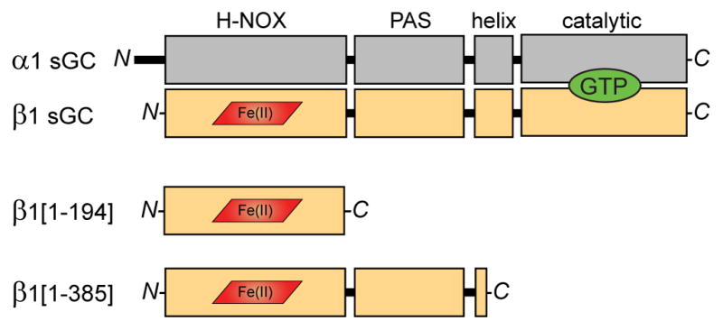
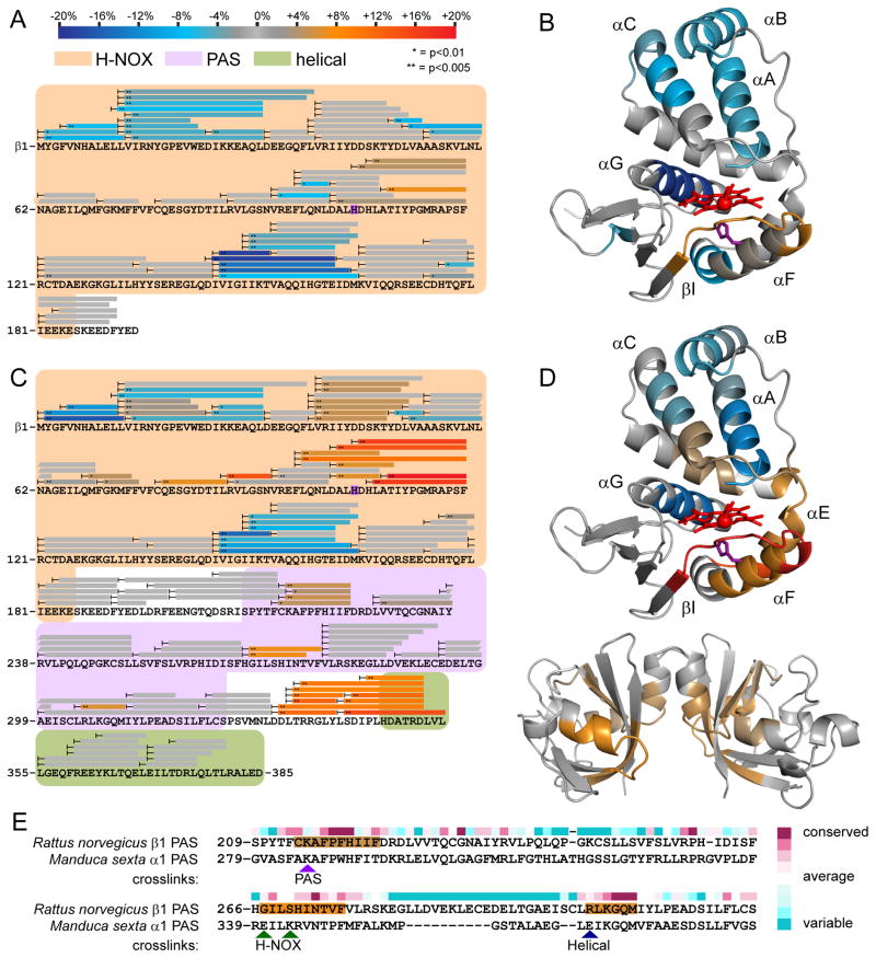
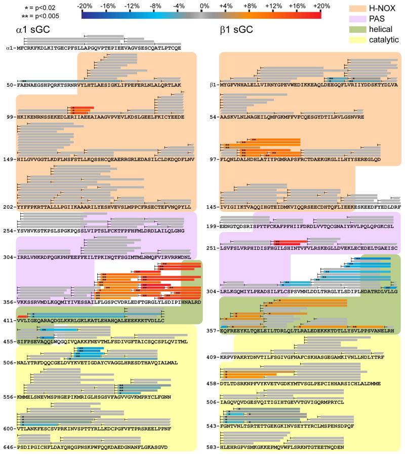
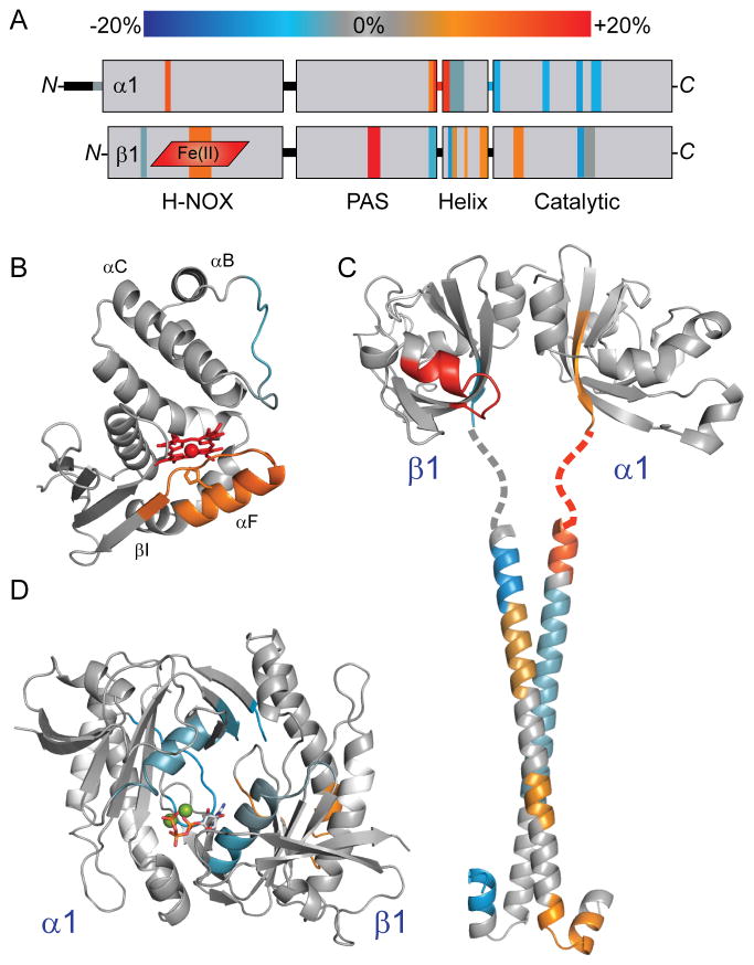
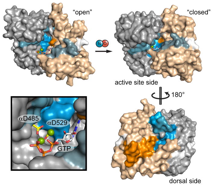
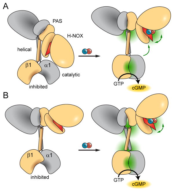
Soluble guanylate cyclase (sGC) is the primary mediator of nitric oxide (NO) signaling. NO binds the sGC heme cofactor stimulating synthesis of the second messenger cyclic-GMP (cGMP). As the central hub of NO/cGMP signaling pathways, sGC is important in diverse physiological processes such as vasodilation and neurotransmission. Nevertheless, the mechanisms underlying NO-induced cyclase activation in sGC remain unclear. Here, hydrogen/deuterium exchange mass spectrometry (HDX-MS) was employed to probe the NO-induced conformational changes of sGC. HDX-MS revealed NO-induced effects in several discrete regions. NO binding to the heme-NO/O2-binding (H-NOX) domain perturbs a signaling surface implicated in Per/Arnt/Sim (PAS) domain interactions. Furthermore, NO elicits striking conformational changes in the junction between the PAS and helical domains that propagate as perturbations throughout the adjoining helices. Ultimately, NO binding stimulates the catalytic domain by contracting the active site pocket. Together, these conformational changes delineate an allosteric pathway linking NO binding to activation of the catalytic domain.


Figures

Figure 1
Figure 1. Domain organization of sGC subunits and truncations
The sGC holoenzyme consists of two homologous subunits, α1 (gray) and β1 (tan), each composed of four domains. NO binds to the ferrous heme cofactor (red) of the β1 H-NOX domain. Cyclization of GTP (green) to cGMP occurs in the C-terminal catalytic domains. Truncations of the β1 sGC subunit preserve NO-binding. β1[1-194] comprises the minimal heme-binding H-NOX domain. β1[1-385] includes the associated PAS domain and a portion of the helical domain.
Figure 2
Figure 2. NO-induced conformational changes of sGC H-NOX domain constructs
Peptide coverage from HDX-MS analysis is represented by bars overlaying sequence. Peptides exhibiting significant NO-induced exchange rate changes are color-coded according to the scale bar. Exchange rate changes are reported as the averaged difference in percent deuterium incorporation (Δ%D) over the actively exchanging portion of the time course. Asterisks denote the results of two-tailed, unpaired Student’s t-tests. Peptides exhibiting non-significant or undetectable changes are colored gray. (A) Changes in HD exchange kinetics induced by NO-binding to β1[1-194]. (B) Color-coded HDX-MS results for NO-induced changes in β1[1-194] were mapped to a homology model derived from the structure of a bacterial H-NOX (PDB: 2o09) (Ma et al., 2007). Heme (red) and the axial heme ligand His-105 (purple) are represented as sticks. (C) Changes in HD exchange kinetics induced by NO-binding to β1[1-385]. (D) Color-coded HDX-MS results for NO-induced changes in β1[1-385] were mapped to the sGC β1 H-NOX model (top) and a β1 PAS domain homodimer model (bottom). The β1 PAS domain was modeled using the Robetta server and dimerized by alignment to the structure of a homologous bacterial PAS dimer (PDB: 2P04) (Kim et al., 2004; Ma et al., 2008). (E) Comparison of NO-induced PAS domain changes with inter-domain contacts identified by crosslinking. Crosslinks identified between the α1 PAS domain of M. sexta sGC and the indicated domains are highlighted with arrowheads(Fritz et al., 2013). NO-induced perturbations identified by HDX-MS are color-coded as in (A) and mapped to the sequence of the rat β1 PAS. Residue conservation (above sequence) of β1 sGC was scored and color-coded using the ConSurf server (Ashkenazy et al., 2010).
Figure 3
Figure 3. NO-induced conformational changes of the full-length sGC heterodimer
Changes in HD exchange kinetics induced by NO-binding in full length α1β1 sGC with noncyclizable GTP analog GPCPP. Peptide coverage from HDX-MS analysis is represented by bars overlaying primary sequence of α1 (left) and β1 (right) sGC. Peptides exhibiting significant NO-induced exchange rate changes are color-coded according to the scale bar. Peptides exchanging more rapidly upon NO-binding are colored in red hues, whereas peptides exchanging more slowly are in blue hues. Asterisks denote the results of two-tailed, unpaired Student’s t-tests. Peptides exhibiting non-significant or undetectable changes are colored gray.
Figure 4
Figure 4. NO-induced conformational changes of full-length sGC mapped to domain structural models
(A) Results from HDX-MS analysis of the NO-induced changes in α1β1 sGC (Figure 3) were condensed, color-coded according to the scale bar, and mapped to the representation of sGC domain organization. (B) HD exchange rate perturbations induced by NO-binding were color-coded (as in Figure 3) and mapped to a structural model of the β1 H-NOX, as in Figure 2. (C) NO-induced changes were mapped to a model of the α1β1 PAS-linker-helical domains. The α1 and β1 PAS domains were modeled independently using the Robetta server, based on homology with the M. sexta α1 PAS domain structure (PDB: 4GJ4) (Kim et al., 2004; Purohit et al., 2013). PAS domain monomers were dimerized by alignment with the structure of the bacterial PAS dimer (PDB: 2P04) (Ma et al., 2008). The structure of the rat sGC β1 helix (PDB: 3HLS) was used to reconstruct the coiled-coil of the helical domain (Ma et al., 2010). The α1 helical subunit was modeled using the Robetta server. The α1β1 coiled-coil was docked using the ClusPro 2.0 server, selecting the highest populated class exhibiting parallel orientation (Comeau et al., 2004). The intervening linker sequence is represented as a dotted line. (D) NO-induced changes were mapped to the structure of the sGC α1β1 catalytic domain heterodimer (PDB: 3UVJ) (Allerston et al., 2013). For reference, GTP:Mg2+ substrate (sticks and balls) was docked into the active site.
Figure 5
Figure 5. Mapping NO-induced conformational changes to models of catalytic domain activation
Catalytic domain exchange rate differences induced by NO were mapped to models of the inactive, “open” conformation and active “closed” conformation. Exchange rate differences are color-coded as in Figure 3; here, α1 and β1 subunits are distinguished as gray and tan, respectively. The open conformation is represented by the structure of the human sGC catalytic domain (PDB: 3UVJ) with GTP docked. The closed conformation is a model based on the structure of a putatively active adenylate cyclase (PDB: 1AZS). Active site aspartate residues, α1 Asp-485 and α1 Asp-529, are highlighted (yellow) in the inset.
Figure 6
Figure 6. Proposed NO-induced activation mechanisms of sGC
NO-binding releases and opens the heme-associated helix of the H-NOX domain while and condensing and closing the active site pocket of the catalytic domain. Dominant points of conformational articulation are highlighted in green. Higher-order domain architecture is based on an emerging single particle electron microscopy study (Campbell MG, ESU, Potter CS, Carragher B, MAM, submitted). The allosteric pathway bridging the sensor and output domains may involve two different mechanisms. (A) Large conformational changes at the junction between PAS and helical domains may indicate inter-domain pivoting that relieves inhibitory contacts between H-NOX and catalytic domains. (B) Alternatively, the PAS—helical junction may mediate remote allosteric effects via long-range conformational changes propagated through the helical domains.

Comment in

References

    1. Allerston CK, von Delft F, Gileadi O. Crystal structures of the catalytic domain of human soluble guanylate cyclase. PLoS One. 2013;8:e57644. - PMC - PubMed
    1. Anantharaman V, Balaji S, Aravind L. The signaling helix: a common functional theme in diverse signaling proteins. Biol Direct. 2006;1 - PMC - PubMed
    1. Ashkenazy H, Erez E, Martz E, Pupko T, Ben-Tal N. ConSurf 2010: calculating evolutionary conservation in sequence and structure of proteins and nucleic acids. Nucleic Acids Res. 2010;38:W529–W533. - PMC - PubMed
    1. Bredt DS. Endogenous nitric oxide synthesis: Biological functions and pathophysiology. Free Radic Res. 1999;31:577–596. - PubMed
    1. Busker M, Neidhardt I, Behrends S. Nitric Oxide Activation of Guanylate Cyclase Pushes the α1 Signaling Helix and the β1 Heme Binding Domain closer to the Substrate Binding Site. J Biol Chem 2013 - PMC - PubMed

Publication types

MeSH terms