. 2014 Mar 6;53(5):689-99.
doi: 10.1016/j.molcel.2014.01.012. Epub 2014 Feb 20.

Polyphosphate is a primordial chaperone

Affiliations

Polyphosphate is a primordial chaperone

Michael J Gray et al. Mol Cell. .

Abstract

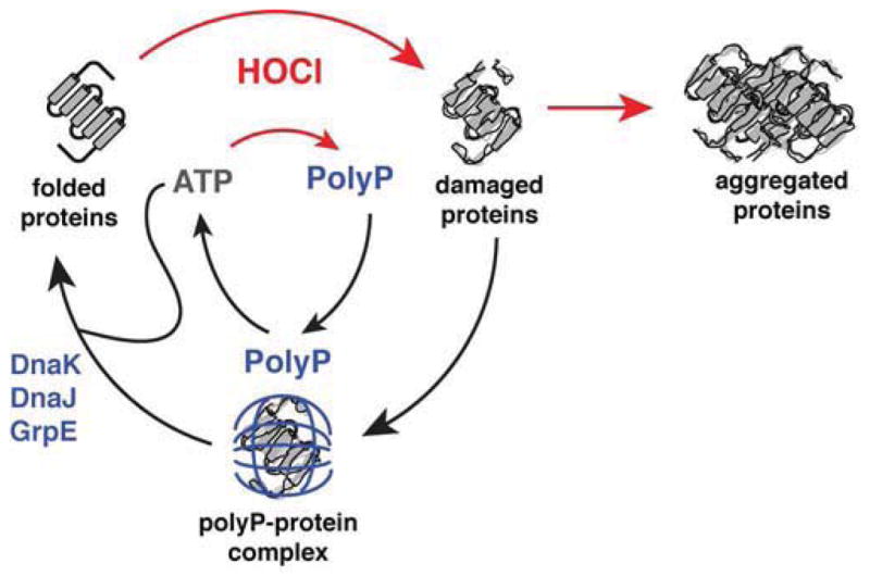
Composed of up to 1,000 phospho-anhydride bond-linked phosphate monomers, inorganic polyphosphate (polyP) is one of the most ancient, conserved, and enigmatic molecules in biology. Here we demonstrate that polyP functions as a hitherto unrecognized chaperone. We show that polyP stabilizes proteins in vivo, diminishes the need for other chaperone systems to survive proteotoxic stress conditions, and protects a wide variety of proteins against stress-induced unfolding and aggregation. In vitro studies reveal that polyP has protein-like chaperone qualities, binds to unfolding proteins with high affinity in an ATP-independent manner, and supports their productive refolding once nonstress conditions are restored. Our results uncover a universally important function for polyP and suggest that these long chains of inorganic phosphate may have served as one of nature's first chaperones, a role that continues to the present day.


Figures

Figure 1
Figure 1. ATP-Derived PolyP Protects Against HOCl
(A) Exponentially growing E. coli wild-type and mutant strains were incubated with 2 mM HOCl for the indicated time, serially diluted, and spot-titered. See also Figures S1A and S1B. (B) Growth (lower panel) and intracellular polyP concentration (upper panel) of E. coli wild-type (black circles), Δppk (red squares), and Δppx (blue triangles) (mean ± SD). Arrow indicates addition of 1 mM HOCl. See also Figures S1C, S1D, and S1E. (C) PolyP synthesis and degradation pathway in E. coli. (D) ATP content of log phase E. coli wild-type (black squares) and Δppk (red circles) with (closed symbols) or without (open symbols) treatment with 1 mM HOCl, as a percentage of the initial value for each sample (mean ± SD). See also Figure S1F. (E) Exponentially growing E. coli wild-type and mutant strains were incubated with 2.5 mM HOCl, then diluted and spot-titered. See also Figures S1G and S1H.
Figure 2
Figure 2. PolyP is a Protein-Protective Chaperone In Vivo
(A) and (B) Wild-type (black) and Δppk (red) E. coli were grown to log phase, then treated with 0.4 mM HOCl. Expression of (A) the heat shock genes ibpA, hslO, dnaK, rpoH, or (B) the DNA damage indicator gene sulA was measured by qRT-PCR (mean ± SD). See also Figure S2A. (C) Insoluble protein fractions from exponentially-growing E. coli wild-type and mutant strains before and after addition of 1 mM HOCl. (D) Survival of (left panels) and insoluble protein fractions from (right panels) exponentially-growing E. coli rpoH strains before and after a shift from 30° to 46°C. (E) E. coli strains containing no polyP (red squares), wild-type (black circles), or higher than normal levels of polyP (blue triangles) were grown to log phase, serially diluted, spot-titered on agar containing different concentrations of spectinomycin and scored for growth (mean ± SD). See also Figures S2B and S2C.
Figure 3
Figure 3. PolyP is a Protein-Protective Chaperone In Vitro
(A) Aggregation of urea-denatured luciferase upon its dilution (arrow) into buffer containing 0 (black), 0.5 (red), 5 (green), 50 (purple), or 500 μM (blue) polyP. PolyP concentrations are expressed in terms of concentration of inorganic phosphate equivalents (Pi). (B) Thermal aggregation of luciferase upon its dilution into pre-warmed buffer (arrow) containing 0 (black), 1 (red), 10 (purple), or 100 μM (blue) polyP. (C) Thermal aggregation of luciferase upon its dilution into pre-warmed buffer containing 0 (black) or 0.5 mM polyP (blue, green, red) and 50 μM MgCl2. Arrow indicates addition of 5 (blue) or 1 (green) μg ml−1 ScPPX. See Figure S3A for additional controls. (D) Circular dichroism spectra of luciferase incubated with or without 0.3 mM polyP at 20° or 85°C. Inset shows SDS-PAGE of soluble and insoluble luciferase fractions after 20 min incubation at the indicated temperature and spectrum determination. (E) Left panel: thermal inactivation of luciferase with 0 (black), 0.1 (blue), 1 (green), or 10 mM polyP (red) at 40°C. Right panel: reactivation of luciferase thermally inactivated in the absence (black) or presence (green) of 1 mM polyP upon shift to permissive temperatures and addition of DnaK, DnaJ, GrpE, and MgATP (KJE). Inset shows reactivation in the absence of KJE. Error bars indicate mean ± SD. (F) Thermal or (G) HOCl-induced aggregation of citrate synthase with 0 (black), 1 (red), 10 (purple), or 100 mM (blue) polyP. Arrow indicates time of citrate synthase addition (in F) or HOCl addition (in G). (H) Crude lysates of E. coli ppk::kan+ were incubated 30 minutes at 30° or 55°C, with 0, 0.2, 1, 2, 10, or 20 mM polyP. Soluble and insoluble fractions were separated and examined by SDS-PAGE. See Figure S3B for results with wild-type lysates.
Figure 4
Figure 4. PolyP Chain Length Influences Chaperone Activity
(A) Crude lysates of E. coli ppk::kan+ were incubated 30 minutes at 30° or 55°C, with 0 or 5 mM of different length polyP; from left to right: heterogeneous short-chain (average 45-mer), 14-mer, 60-mer, 130-mer, and long-chain (200–1,300-mer). Insoluble fractions were separated and examined by SDS-PAGE. (B) and (C) Aggregation of urea-denatured citrate synthase (B) or thermally-denatured luciferase (C) upon their dilution into buffer (arrow) containing no polyP (black) or the indicated concentrations of different length polyP: 14-mer (blue), 60-mer (green), 130-mer (orange), or 300-mer (red). Concentrations were determined according to the length of polyP chains. Corresponding experiments using total Pi concentration are shown in Figures S4A and S4B.
Figure 5
Figure 5. PPX is a Redox-Regulated Enzyme
(A) Specific activity of PPX after incubation with different concentrations of N-chlorotaurine (NCT) (mean ± SD). To test for reversibility, oxidatively inactivated PPX (NCT:PPX 10:1) was incubated for 1 hr with 5 mM DTT and assayed again. See also Figure S4A. (B) Specific activity of PPX after treatment with H2O2 (mean ± SD). (C) E. coli overexpressing PPX was grown to log phase, then treated with 1 mM HOCl. Reduced cysteine thiols were alkylated with iodoacetamide (IAM). Oxidized cysteine thiols were reduced and alkylated with PEG-maleimide (PEG), adding 2 kDa molecular mass per modified cysteine. PPX was visualized by Western blot. Fully IAM-labeled (“Reduced”) or PEG-maleimide-labeled (“Oxidized”) PPX standards are from the same blot, with longer exposure times. (D) and (E) Specific activity of PPK’s forward and reverse reactions (see Figure 1C) before and after treatment with NCT (mean ± SD). See also Figure S4B.
Figure 6
Figure 6. Model of the PolyP Chaperone Cycle
The antimicrobial oxidant HOCl damages proteins, causing formation of cytotoxic protein aggregates. HOCl stimulates rapid conversion of cellular ATP to polyP through the oxidative inactivation of PPX. This conversion conserves high-energy phospho-anhydride bonds while down-regulating cellular processes that require ATP, including ATP-dependent chaperones like DnaK. PolyP functionally replaces these chaperones by forming stable complexes with unfolding proteins, keeping them soluble and refolding-competent. Upon relief of stress conditions, polyP may be either degraded to free phosphate by PPX or reconverted to ATP by PPK. Restoration of cellular ATP pools reactivates ATP-dependent chaperones and allows for the effective refolding of polyP-protected proteins by the DnaK/DnaJ/GrpE complex.

Comment in

References

    1. Achbergerova L, Nahalka J. Polyphosphate--an ancient energy source and active metabolic regulator. Microb Cell Fact. 2011;10:63. - PMC - PubMed
    1. Alvarado J, Ghosh A, Janovitz T, Jauregui A, Hasson MS, Sanders DA. Origin of exopolyphosphatase processivity: Fusion of an ASKHA phosphotransferase and a cyclic nucleotide phosphodiesterase homolog. Structure. 2006;14:1263–1272. - PubMed
    1. Anfinsen CB. Principles that govern the folding of protein chains. Science. 1973;181:223–230. - PubMed
    1. Antelmann H, Helmann JD. Thiol-based redox switches and gene regulation. Antioxidants & redox signaling. 2011;14:1049–1063. - PMC - PubMed
    1. Aschar-Sobbi R, Abramov AY, Diao C, Kargacin ME, Kargacin GJ, French RJ, Pavlov E. High sensitivity, quantitative measurements of polyphosphate using a new DAPI-based approach. J Fluoresc. 2008;18:859–866. - PubMed

Publication types

MeSH terms