. 2014 Mar 13;6(5):844-54.
doi: 10.1016/j.celrep.2014.01.030. Epub 2014 Feb 20.

PTEN C-terminal deletion causes genomic instability and tumor development

Affiliations

PTEN C-terminal deletion causes genomic instability and tumor development

Zhuo Sun et al. Cell Rep. .

Abstract

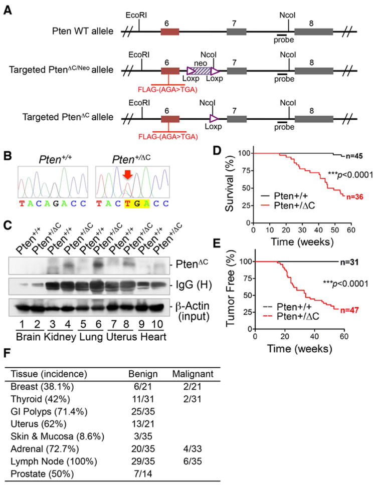
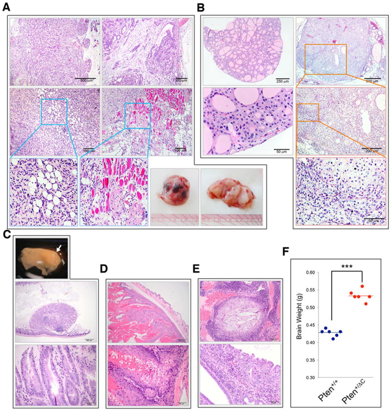
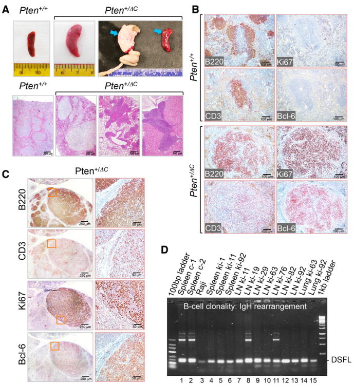
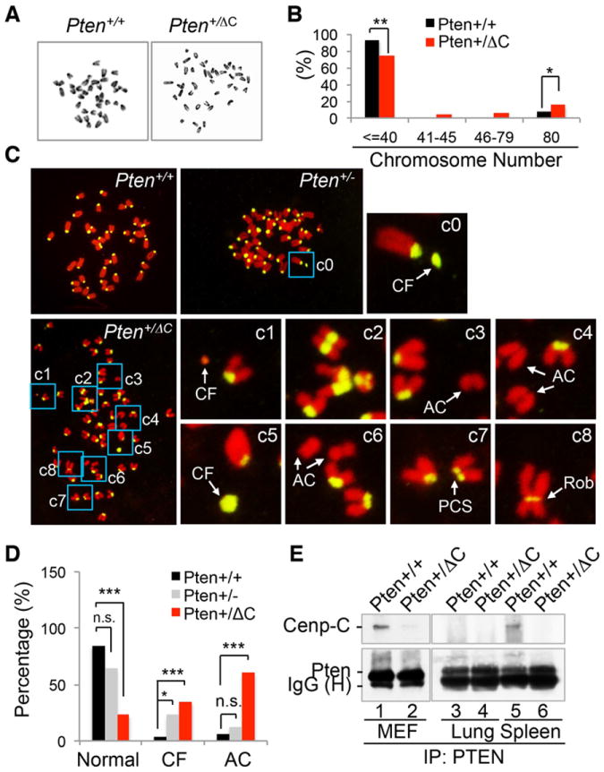
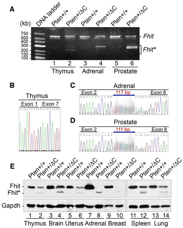
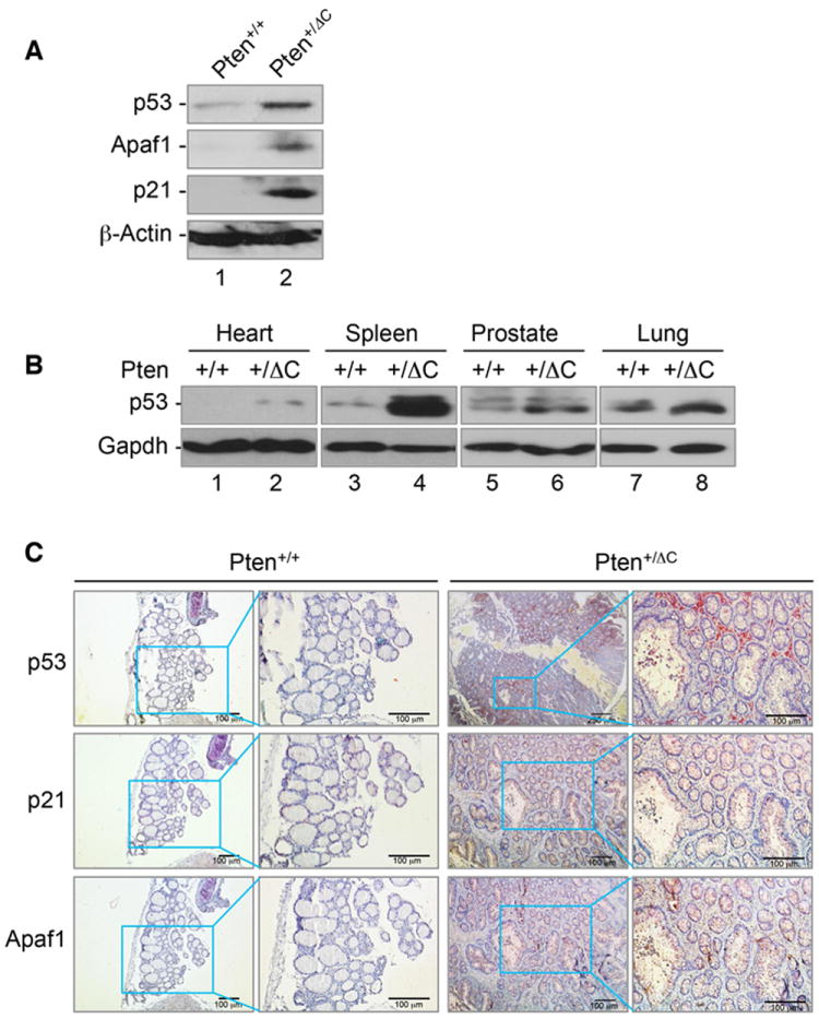
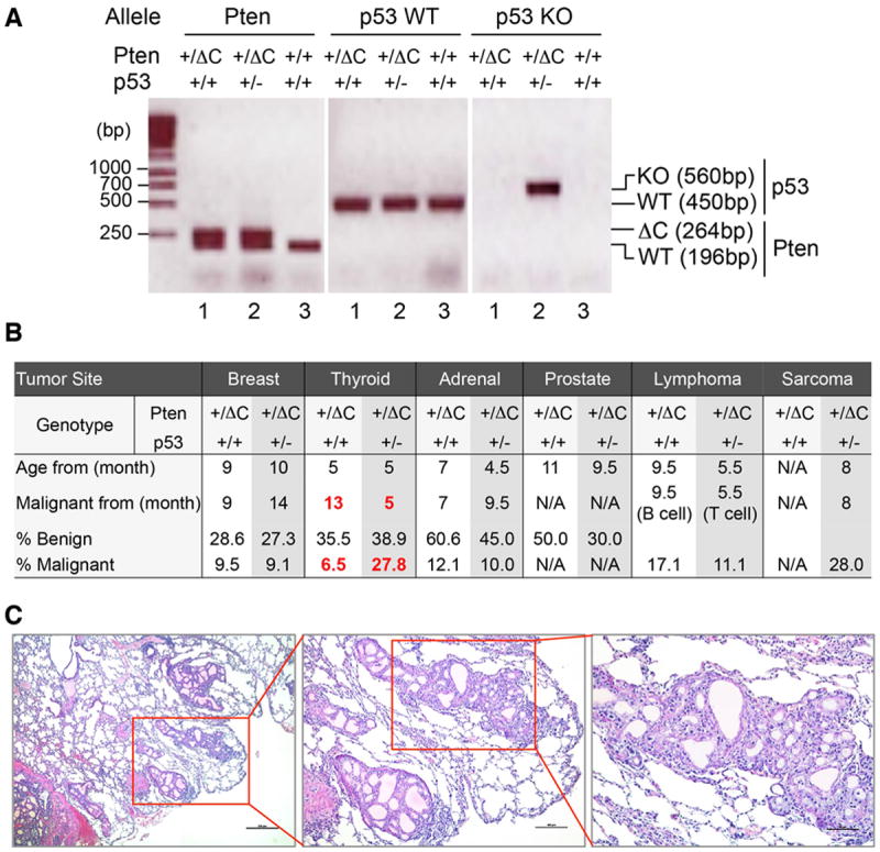
Tumor suppressor PTEN controls genomic stability and inhibits tumorigenesis. The N-terminal phosphatase domain of PTEN antagonizes the PI3K/AKT pathway, but its C-terminal function is less defined. Here, we describe a knockin mouse model of a nonsense mutation that results in the deletion of the entire Pten C-terminal region, referred to as Pten(ΔC). Mice heterozygous for Pten(ΔC) develop multiple spontaneous tumors, including cancers and B cell lymphoma. Heterozygous deletion of the Pten C-terminal domain also causes genomic instability and common fragile site rearrangement. We found that Pten C-terminal disruption induces p53 and its downstream targets. Simultaneous depletion of p53 promotes metastasis without influencing the initiation of tumors, suggesting that p53 mainly suppresses tumor progression. Our data highlight the essential role of the PTEN C terminus in the maintenance of genomic stability and suppression of tumorigenesis.


Figures

Figure 1
Figure 1. Knock-in of C-terminal truncated Pten reduces mouse lifespan due to spontaneous tumor development
(A) Schematic diagram of strategy for knock-in of the Pten point mutation, c.565A>T, leading to deletion of the Pten C-terminal domain (aa189-403). (B) Confirmation of the point mutation of c.565A>T by direct sequencing of Pten exon 6 in Pten+/+ and Pten+/ΔC ES clones. (C) The expression of the C-terminal truncated PtenΔC in various tissues of Pten+/ΔC mice examined by immunoprecipitation with an N-terminus-specific PTEN antibody prior to FLAG immunoblotting. Corresponding tissues from wild-type mice were included as controls. (D) Kaplan-Meier plot for overall survival of Pten+/+ and Pten+/ΔC mice. (E) Kaplan-Meier plot showing significantly shorter latency of tumor development in Pten+/ΔC mice as compared with wild-type control mice. (F) Incidence of tumor in different tissues. Pten+/ΔC mice aged from 12 to 60 weeks (n=35, 14 males and 21 females) were subjected to extensive histological evaluation. Enlarged lymph nodes were found in every mouse analyzed. Pheochromocytoma occurred in all Pten+/ΔC mice older than six months with a 12% frequency of pulmonary metastasis. See also Figure S1.
Figure 2
Figure 2. Pten+/ΔC mice exhibit Cowden syndrome-like tumors in multiple tissues
(A) Benign and malignant mammary tumors. Top panels, complex mammary adenoma; Middle and bottom left panels, carcinoma with areas magnified to show infiltrative growth of adipose tissue and skeletal muscle; Bottom right panel, gross view of a breast tumor with multiple foci of hemorrhagic necrosis. (B) Follicular adenoma of the thyroid (left panels) and thyroid carcinoma (right panels). The carcinoma consists of a large expansile nodule. Selected areas of the tumor are magnified to show follicular cells arrayed in disorderly, multilobulated nodules with atypia and very little follicle formation reflecting lack of differentiation in this carcinoma. (C) A polyp of the large intestine showing features of a hamartomatous polyp shown in gross view (top, white arrow) and histological views (middle and bottom). (D) Sebaceous adenoma of the skin consisting of a pseudoencapsulated expansile neoplasm showing features of skin appendage architecture. (E) Endometrial epithelial hyperplasia with dysplasia showing endometrium expanded by moderate cystic dilation of endometrial glands and disorderly growth of epithelial cells. (F) Macrocephaly manifested by significantly increased brain weight in Pten+/ΔC mice as compared with age- and gender-matched wild-type control mice. ***, p<0.0001. See also Figure S2.
Figure 3
Figure 3. Pten+/ΔC mice develop lymphoma predominantly of B cell lineage
(A) Splenomegaly and lymphoma. The upper panel shows gross views of enlarged spleen (arrows point to a mesenteric lymph node and nodular aggregates in the spleen) in Pten+/ΔC mice. The lower panel displays histological images of abnormal splenic architecture (lower mid-left) and lymphoma in the lung (lower mid-right) and kidney (lower right). Normal spleen from a wild-type mouse is shown (lower left) as a control. (B) Lymphoma in the spleen from a Pten+/ΔC mouse. Immunohistochemical analysis of B (B220), T (CD3), proliferation (Ki67) cell markers and Bcl-6 in spleens from Pten+/+ and Pten+/ΔC mice. (C) B cell lymphoma with strong expression of the Bcl-6 marker. Lymph nodes from Pten+/ΔC mice were subjected to immunohistochemical staining for indicated cell markers. (D) Validation of B cell lymphoma lineage derivation by clonality analysis. Clonal B cell expansion is identified by monotonous D-J rearrangement of IgH in 10 out of 12 biopsies (lanes 3-7, 9, 10, 12-15). See also Figure S4.
Figure 4
Figure 4. Knock-in of PtenΔC causes chromosomal instability
(A and B) Aneuploidy and polyploidy. Chromosome number was examined in metaphase spreads prepared from Pten+/+ (n=101) and Pten+/ΔC (n=126) MEFs (A) and data are summarized in the histogram (B). Images shown above the histogram are representative metaphase spreads. *, p<0.05; **, p<0.01. (C) Chromosomal aberrations featured by centromeric abnormalities. Metaphase spreads from Pten+/+ (n=32), Pten+/- (n=48) and Pten+/ΔC (n=43) MEFs were hybridized with a pan-centromeric probe (yellow) and DNA was stained with propidium iodide. Areas with aberrant chromosomes are magnified for visual enhancement. CF, centromeric fragment; AC, acentric chromosome fragment; PCS, premature centromeric separation; Rob, Robertsonian chromosomal fusion. (D) A summary of frequencies of centromeric fragments (CF, >2 per cell) and acentric chromosomes (AC). (E) Loss of physical association with CENP-C. Protein lysates from Pten+/+ and Pten+/ΔC cells or tissues were immunoprecipitated with a PTEN monoclonal antibody followed by immunoblotting with a rabbit anti-CENP-C antibody. See also Figure S5.
Figure 5
Figure 5. Knock-in of PtenΔC results in common fragile site instability
(A-D) Aberrant transcripts of Fhit in different tissues from Pten+/ΔC mice due to alternative splicing and translocation. Transcripts of Fhit were amplified with nested PCR from cDNA of indicated tissues (A) and abnormal transcripts were purified for sequencing (B-D). (E) Reduction of normal Fhit expression (~17 kDa) and production of a smaller Fhit protein variant (~15 kDa). Various pairs of tissues from Pten+/+ and Pten+/ΔC mice as indicated were analyzed for Fhit expression by Western blot. See also Figure S6 and Table S1.
Figure 6
Figure 6. Pten C-terminal deletion induces the p53 signaling pathway
(A) Upregulation of p53 and its downstream targets in Pten+/ΔC MEFs as compared with wild type cells. Western blot analysis of p53 and its target genes in Pten+/+ and Pten+/ΔC MEFs using specific antibodies as indicated. (B) p53 induction in various tissues lacking the Pten C-terminal region. Various tissues from mice with and without Pten C-terminal deletion were subjected to immunoblotting analysis of p53 expression. (C) Thyroid tissues from Pten+/+ and Pten+/ΔC mice were stained by immunohistochemistry for p53, p21 and Apaf1, showing increased signals in PtenΔC knock-in tissues.
Figure 7
Figure 7. Inactivation of p53 in Pten+/ΔC mice facilitates malignancy and promotes metastasis of thyroid tumors
(A) Generation of double knockout mice with heterozygous p53 and PtenΔC alleles. Genotyping PCR analysis of tail DNA from Pten+/+ p53+/+, Pten+/ΔC p53+/+ and Pten+/ΔC p53+/- mice. (B) A summary of tumor incidence based on extensive histological evaluation. (C) Representative histological images showing thyroid tumor metastasis in lung.

References

    1. Anzelon AN, Wu H, Rickert RC. Pten inactivation alters peripheral B lymphocyte fate and reconstitutes CD19 function. Nat Immunol. 2003;4:287–294. - PubMed
    1. Aqeilan RI, Trapasso F, Hussain S, Costinean S, Marshall D, Pekarsky Y, Hagan JP, Zanesi N, Kaou M, Stein GS, et al. Targeted deletion of Wwox reveals a tumor suppressor function. Proc Natl Acad Sci U S A. 2007;104:3949–3954. - PMC - PubMed
    1. Basso K, Dalla-Favera R. BCL6: master regulator of the germinal center reaction and key oncogene in B cell lymphomagenesis. Adv Immunol. 2010;105:193–210. - PubMed
    1. Bubien V, Bonnet F, Brouste V, Hoppe S, Barouk-Simonet E, David A, Edery P, Bottani A, Layet V, Caron O, et al. High cumulative risks of cancer in patients with PTEN hamartoma tumour syndrome. J Med Genet. 2013;50:255–263. - PubMed
    1. Cattoretti G, Pasqualucci L, Ballon G, Tam W, Nandula SV, Shen Q, Mo T, Murty VV, Dalla-Favera R. Deregulated BCL6 expression recapitulates the pathogenesis of human diffuse large B cell lymphomas in mice. Cancer Cell. 2005;7:445–455. - PubMed

Publication types

MeSH terms

Substances

LinkOut - more resources