. 2014 Mar 4;19(3):498-511.
doi: 10.1016/j.cmet.2014.02.001. Epub 2014 Feb 20.

Islet microenvironment, modulated by vascular endothelial growth factor-A signaling, promotes β cell regeneration

Affiliations

Islet microenvironment, modulated by vascular endothelial growth factor-A signaling, promotes β cell regeneration

Marcela Brissova et al. Cell Metab. .

Erratum in

  • Cell Metab. 2015 Oct 6;22(4):750

Abstract

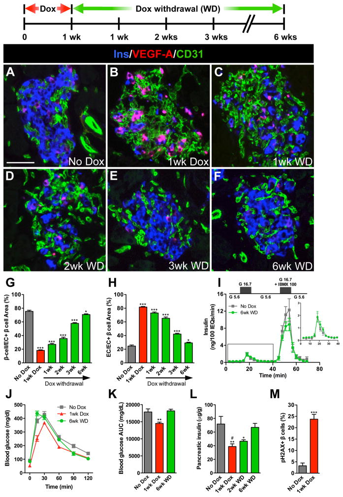
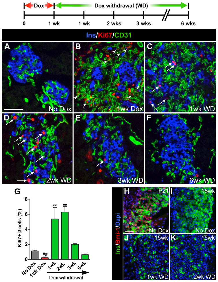
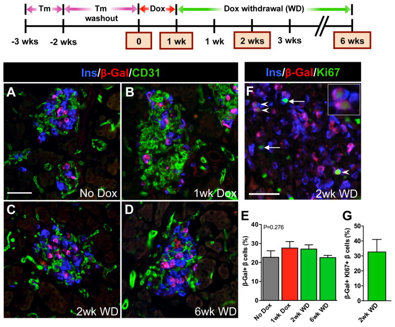
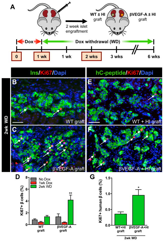
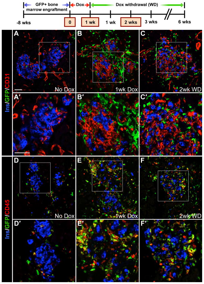
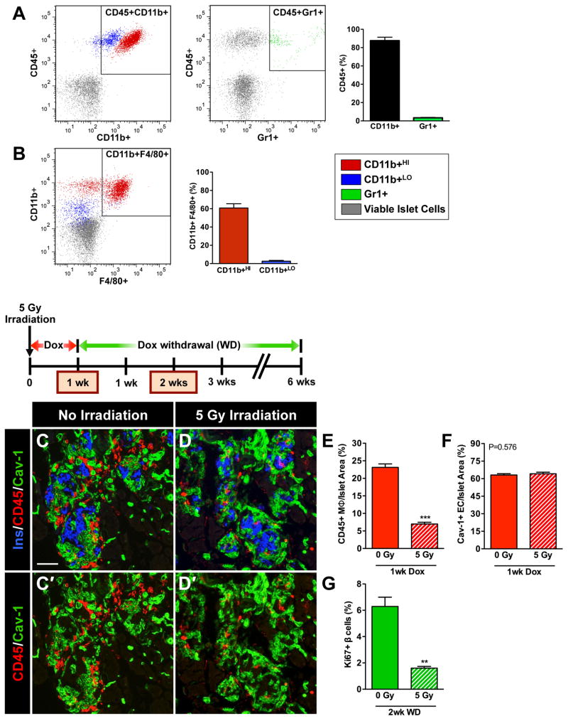
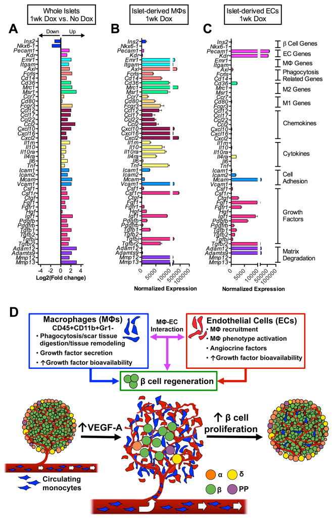
Pancreatic islet endocrine cell and endothelial cell (EC) interactions mediated by vascular endothelial growth factor-A (VEGF-A) signaling are important for islet differentiation and the formation of highly vascularized islets. To dissect how VEGF-A signaling modulates intra-islet vasculature, islet microenvironment, and β cell mass, we transiently increased VEGF-A production by β cells. VEGF-A induction dramatically increased the number of intra-islet ECs but led to β cell loss. After withdrawal of the VEGF-A stimulus, β cell mass, function, and islet structure normalized as a result of a robust, but transient, burst in proliferation of pre-existing β cells. Bone marrow-derived macrophages (MΦs) recruited to the site of β cell injury were crucial for the β cell proliferation, which was independent of pancreatic location and circulating factors such as glucose. Identification of the signals responsible for the proliferation of adult, terminally differentiated β cells will improve strategies aimed at β cell regeneration and expansion.


Figures

Figure 1
Figure 1. Increasing β cell VEGF-A production increases intra-islet ECs but leads to β cell loss followed by β cell regeneration after withdrawal of the VEGF-A stimulus
VEGF-A was induced for 1 wk by Dox administration followed by 6 wks of Dox withdrawal (WD). (A–F) Labeling for insulin (Ins, blue), VEGF-A (red), and EC marker CD31 (green). Scale bar is 50 μm and applies to A–F. (G, H) Relationship between β cells (G) and intra-islet ECs (H) upon induction and withdrawal of the VEGF-A stimulus. ***, p<0.001, 1wk Dox, 1wk WD, 2wk WD, or 3wk WD vs. No Dox control; *, p<0.05, 6wk WD vs. No Dox control; n=4 mice/time point. Islet size measured by pixel area increased slightly but not significantly with intra-islet EC expansion; No Dox, 6545±1687 pixels; 1wk Dox, 10705±1712 pixels, p=0.1939. (I) Islets isolated from No Dox controls and at 6wk WD (n=4 mice/group) were examined in a cell perifusion system. Both groups had normal basal insulin secretion at 5.6 mM glucose (G 5.6) and the magnitude of the insulin secretory response was similar when stimulated with either 16.7 mM glucose (G 16.7; 9.6±2.1 vs. 11.3±2.5 ng/100 IEQ, p=0.63) or 16.7 mM glucose + 100 μM IBMX (G 16.7 + IBMX 100; 111±22 vs. 84±13 ng/100 IEQ, p=0.34). Inset shows enlarged boxed portion of insulin secretory profile. (J, K) Glucose clearance in βVEGF-A mice. **, p<0.01, 1wk Dox (n=10) vs. No Dox (n=14) or 6wk WD (n=5). (L) Loss in pancreatic insulin content 1 wk after VEGF-A induction was restored over the 6 wks following Dox withdrawal. **, p<0.01, 1wk Dox vs. No Dox; *, p<0.05, 2wk WD vs. No Dox; #, p<0.05, 1wk Dox vs. 6wk WD; No Dox vs. 6wk WD was not statistically significant; n=6–7 mice/time point. (M) Increased β cell apoptosis 1 wk after VEGF-A induction; n=4 mice/time point; 100–500 β cells analyzed/mouse. ***, p<0.001, 1wk Dox vs. No Dox.
Figure 2
Figure 2. Removal of the VEGF-A stimulus results in a transient burst in β cell proliferation
β cell proliferation was monitored during the experimental period outlined; n=4 mice/time point. (A–F) Labeling for insulin (Ins, blue), Ki67 (red), and CD31 (green). Scale bar is 50 μm and applies to A–F. (G) Quantification of β cell proliferation. ##, p<0.01, 1wk Dox vs. No Dox, 1wk WD, 2wk WD, or 3wk WD. **, p<0.01, 1wk WD or 2wk WD vs. No Dox, 1wk Dox, 3wk WD, or 6wk WD. No Dox, 1wk Dox, 3wk WD, and 6wk WD comparisons were not statistically significant. (H–K) Increased β cell proliferation was not associated with increased Bmi–1; insulin (Ins, green), Bmi-1 (red), Dapi (blue). Scale bar is 25 μm and applies to H–K.
Figure 3
Figure 3. New β cells in the βVEGF-A model arise from replication of pre-existing β cells
β cells in RIP-rtTA;Tet-O-VEGF-A;Pdx1PB-CreERTm;R26Rwt/lacZ transgenic mice were genetically labeled by Tm injection 2 wks prior to inducing VEGF-A for 1 wk by Dox administration followed by 6 wks of Dox withdrawal. Expression of β-gal in β cells was analyzed in 3–4 mice/time point. (A–D) Labeling for insulin (Ins, blue), β-galactosidase (β-Gal, red), and CD31 (green). Scale bar is 50 μm and applies to A–D. (E) β-Gal labeling index in β cells was not statistically different at any time point, p=0.2760. (F) Genetically labeled β cells proliferate. Arrowheads denote β-gal+Ki67+ β cells, progeny of surviving and proliferating β cells (enlargement in inset). Arrows point to β-gal-Ki67+ β cells. Scale bar is 50 μm. (G) One-third of all proliferating β cells expressed the β-gal genetic mark, which is consistent with genetic labeling in panel E. The β cell proliferation index at 2wk WD was 6.1±1.0%, consistent with Figure 2G.
Figure 4
Figure 4. β cell replication is independent of the pancreatic site and soluble circulating factors and is not limited to murine β cells
(A) Islets from βVEGF-A mice and WT controls were transplanted into βVEGF-A recipients or mixed with human islets (HI) and transplanted into NOD-scid-IL2rγnull mice. Islets engrafted for 2 wks then grafts were harvested and analyzed at No Dox, 1wk Dox, and 2wk WD time points; n=3–4 mice/time point. (B–C) β cell proliferation at 2wk WD in WT and βVEGF-A islet grafts; insulin (Ins, green), Ki67 (red), and Dapi (blue). Scale bar is 50 μm and applies to B–C. (D) Quantification of β cell proliferation in WT and βVEGF-A islet grafts. **, p<0.01, 2wk WD vs. No Dox and 1wk Dox across graft types. (E–F) β cell proliferation at 2wk WD in WT+HI and βVEGF-A+HI grafts; human C-peptide (hC-peptide, green), Ki67 (red), and Dapi (blue). Scale bar is 50 μm and applies to E–F. (G) Quantification of β cell proliferation in WT+HI and βVEGF-A+HI grafts at 2wk WD. *, p<0.05.
Figure 5
Figure 5. CD45+ BMCs are recruited to the site of β cell injury upon VEGF-A induction and persist in islet remnants during β cell regeneration
βVEGF-A mice were transplanted with GFP+ bone marrow, and 8 wks later VEGF-A was induced for 1 wk by Dox administration followed by 2 wks of Dox withdrawal; n=4–6 mice/time point. (A–C) Labeling for insulin (Ins, blue), CD31 (red) and GFP (green). (D–F) Labeling for insulin (Ins, blue), pan-hematopoietic marker CD45 (red) and GFP (green). Boxes in A–F denote enlargements in A′–F′. Scale bar in A is 50 μm and applies to A–F. Scale bar in A′ is 50 μm and applies to A′–F′.
Figure 6
Figure 6. Recruitment of MΦ into βVEGF-A islets upon VEGF-A induction is necessary for the β cell proliferative response
(A–B) Flow cytometry analysis of βVEGF-A islets after 1-wk Dox treatment. (A) The CD45+ BMC population recruited to βVEGF-A islets was composed of 90% CD11b+ MΦs and 3% Gr1+ cells. (B) F4/80 expression in subsets of more mature CD11b+HI (60%) and less mature CD11b+LO (3%) MΦs; n=5 islet preparations (1–2 mice/preparation). (C–G) Partial bone marrow ablation prior to VEGF-A induction blocks MΦ recruitment and prevents β cell proliferation. (C, D) Immediately after sublethal irradiation, VEGF-A expression in βVEGF-A mice was induced for 1 wk by Dox administration and tissues were examined for the presence of CD45+ MΦs at 1wk Dox and compared with non-irradiated controls; n=4 mice/group; insulin (Ins, blue), CD45 (red) and EC marker caveolin-1 (Cav-1, green). Panel C′ and D′ show CD45 (red) and caveolin-1 (green) labeling. Scale bar is 50 μm and applies to C–D′. (E) Sublethal irradiation reduced infiltration of CD45+ MΦs, ***, p<0.001, 0 Gy vs. 5 Gy. (F) Sublethal irradiation did not affect intra-islet EC expansion, p=0.5760, 0 Gy vs. 5 Gy. (G) Two wks after Dox withdrawal, β cell proliferation was significantly reduced in sublethally irradiated βVEGF-A mice vs. non-irradiated controls, **, p<0.01; n=4 mice/group.
Figure 7
Figure 7. Transcriptome analysis of βVEGF-A islets and purified islet-derived MΦs and ECs
(A–C) Gene expression profile of whole βVEGF-A islets, islet-derived MΦs, and islet-derived ECs by RNA-Seq; n=3 replicates/each sample set. (A) Differential expression of β cell-, EC-, MΦ-specific genes, phagocytosis-related genes, MΦ phenotype markers (M1, classical; M2, alternative), chemokines, cytokines, cell adhesion molecules, growth factors, and matrix degrading enzymes between islets at 1wk Dox vs. No Dox, p<0.05 for fold change ≥2. See also data in Figure S6M. (B) At 1wk Dox, recruited MΦs are highly enriched for transcripts of phagocytosis-related genes, M2 markers, chemokines, cytokines, cell adhesion molecules, and metalloproteinases involved in tissue repair. (C) Intra-islet ECs mainly express growth factors and matrix degrading enzymes that facilitate growth factor release from the extracellular matrix. Data in B and C are plotted as mean ± SEM. (D) New paradigm for the role of MΦ – EC interactions in β cell regeneration. Upon VEGF-A induction intra-islet ECs proliferate and circulating monocytes recruited to islets differentiate into MΦs. These CD45+CD11b+Gr1− recruited MΦs and ECs produce effector molecules that either directly or cooperatively induce β cell proliferation and regeneration.

References

    1. Agudo J, Ayuso E, Jimenez V, Casellas A, Mallol C, Salavert A, Tafuro S, Obach M, Ruzo A, Moya M, et al. Vascular Endothelial Growth Factor-Mediated Islet Hypervascularization and Inflammation Contribute to Progressive Reduction of β-Cell Mass. Diabetes. 2012;61:2851–2861. - PMC - PubMed
    1. Apte SS. A disintegrin-like and metalloprotease (reprolysin-type) with thrombospondin type 1 motif (ADAMTS) superfamily: functions and mechanisms. Journal of Biological Chemistry. 2009;284:31493–31497. - PMC - PubMed
    1. Banaei-Bouchareb L, Gouon-Evans V, Samara-Boustani D, Castellotti MC, Czernichow P, Pollard JW, Polak M. Insulin cell mass is altered in Csf1op/Csf1op macrophage-deficient mice. J Leukoc Biol. 2004;76:359–367. - PubMed
    1. Bardin N, Blot-Chabaud M, Despoix N, Kebir A, Harhouri K, Arsanto JP, Espinosa L, Perrin P, Robert S, Vely F, et al. CD146 and its soluble form regulate monocyte transendothelial migration. Arterioscler Thromb Vasc Biol. 2009;29:746–753. - PubMed
    1. Barleon B, Sozzani S, Zhou D, Weich HA, Mantovani A, Marmé D. Migration of human monocytes in response to vascular endothelial growth factor (VEGF) is mediated via the VEGF receptor flt-1. Blood. 1996;87:3336–3343. - PubMed

Publication types

MeSH terms

Substances