. 2014 May;80(9):2773-85.
doi: 10.1128/AEM.00078-14. Epub 2014 Feb 21.

Dimethylglycine provides salt and temperature stress protection to Bacillus subtilis

Affiliations

Dimethylglycine provides salt and temperature stress protection to Bacillus subtilis

Abdallah Bashir et al. Appl Environ Microbiol. 2014 May.

Abstract

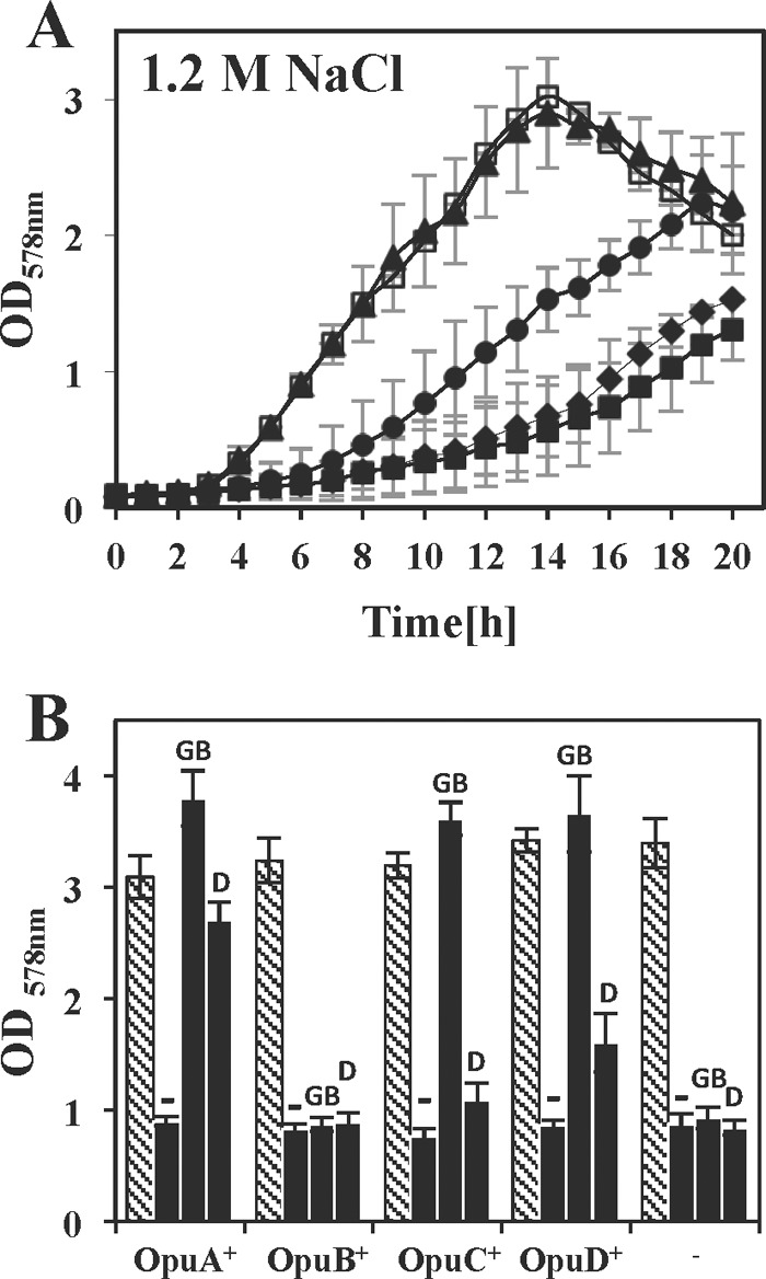
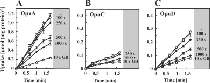
Glycine betaine is a potent osmotic and thermal stress protectant of many microorganisms. Its synthesis from glycine results in the formation of the intermediates monomethylglycine (sarcosine) and dimethylglycine (DMG), and these compounds are also produced when it is catabolized. Bacillus subtilis does not produce sarcosine or DMG, and it cannot metabolize these compounds. Here we have studied the potential of sarcosine and DMG to protect B. subtilis against osmotic, heat, and cold stress. Sarcosine, a compatible solute that possesses considerable protein-stabilizing properties, did not serve as a stress protectant of B. subtilis. DMG, on the other hand, proved to be only moderately effective as an osmotic stress protectant, but it exhibited good heat stress-relieving and excellent cold stress-relieving properties. DMG is imported into B. subtilis cells primarily under osmotic and temperature stress conditions via OpuA, a member of the ABC family of transporters. Ligand-binding studies with the extracellular solute receptor (OpuAC) of the OpuA system showed that OpuAC possesses a moderate affinity for DMG, with a Kd value of approximate 172 μM; its Kd for glycine betaine is about 26 μM. Docking studies using the crystal structures of the OpuAC protein with the sulfur analog of DMG, dimethylsulfonioacetate, as a template suggest a model of how the DMG molecule can be stably accommodated within the aromatic cage of the OpuAC ligand-binding pocket. Collectively, our data show that the ability to acquire DMG from exogenous sources under stressful environmental conditions helps the B. subtilis cell to cope with growth-restricting osmotic and temperature challenges.


Figures

FIG 1
FIG 1
Chemical structures of monomethylglycine (sarcosine), DMG, and glycine betaine.
FIG 2
FIG 2
Protection of B. subtilis against high-salinity growth conditions by compatible solutes. (A) Growth curves of wild-type strain JH642 cultivated at 37°C in SMM with 1.2 M NaCl in the absence (◼) or presence of the solute sarcosine (◆), DMG (●), or glycine betaine (▲) at a final concentration of 1 mM. A culture without 1.2 M NaCl served as an unstressed control (◻). The values shown are the means and standard deviations of three independently grown cultures. (B) Growth yields of cultures grown in the absence (hatched bars) or presence (black bars) of 1.2 M NaCl without (−) or with the addition of 1 mM glycine betaine (GB) or DMG (D). A set of mutants derived from B. subtilis strain JH642 that each express only one or none (−) of the Opu transporters were grown for 20 h at 37°C; the growth yields of the cultures were determined by OD578 measurement. The values shown are the means and standard deviations of two independently grown cultures. The osmotically controlled proline transporter OpuE was present in all of the strains.
FIG 3
FIG 3
Inhibition of [1-14C]glycine betaine by an excess of DMG. B. subtilis mutant strains expressing only the indicated Opu glycine betaine (GB) uptake systems were cultivated in SMM with 1.2 M NaCl. When the cultures reached the early exponential growth phase (OD578 of about 0.3), 2-ml aliquots were withdrawn and immediately mixed with a solution containing a mixture of nonlabeled glycine betaine and [1-14C]glycine betaine (the final concentration in the assay was 10 μM), and the uptake of glycine betaine (○) was then monitored over time. Inhibition of glycine betaine uptake was tested in parallel assays with excesses of DMG (100-, 250-, 500-, and 1,000-fold) as indicated (●). As a control, a 10-fold excess of unlabeled glycine betaine was added to the labeled glycine betaine standard mixture (△) to monitor the inhibition of [1-14C]glycine betaine import by glycine betaine itself. The values shown are the means and standard deviations of three independent cultures.
FIG 4
FIG 4
Protection of B. subtilis growth against high-temperature challenges. (A, C) Growth curves of wild-type strain JH642 cultivated in SMM at 52°C (A) or at the upper growth limit of 52.2°C (C) in the absence (□) or presence of the solute monomethylglycine (◆), DMG (●), or glycine betaine (◼) at a final concentration of 1 mM. The values shown are the means and standard deviations of three independently grown cultures. (B) Growth yields of cultures grown in SMM at 52°C in the absence (hatched bars) or presence of 1 mM DMG (black bars) or glycine betaine (gray bars). A set of B. subtilis mutants derived from strain JH642 that each express only one or none (−) of the relevant Opu transporters were grown for 14 h at 52°C; the growth yields of the cultures were determined by OD578 measurement. The values shown are the means and standard deviations of two independently grown cultures. The osmotically controlled proline transporter OpuE was present in all of the strains.
FIG 5
FIG 5
Protection of B. subtilis growth against low-temperature challenges. (A) Growth curves of B. subtilis wild-type strain 168 cultivated in SMM at 13°C in the absence (◻) or presence of the solute sarcosine (◆), DMG (●), or glycine betaine (◼) at a final concentration of 1 mM. The values shown are the means and standard deviations of three independently grown cultures. (B) Growth yields of cultures grown in SMM at 13°C in the absence (hatched bars) or presence of 1 mM DMG (black bars) or glycine betaine (gray bars) after 10 days of incubation as determined by OD578 measurement. A set of B. subtilis strain 168-derived mutants that each express only one or none (−) of the relevant Opu transporters were used for this experiment. The values shown are the means and standard deviations of two independently grown cultures. The osmotically controlled proline transporter OpuE was present in all of the strains.
FIG 6
FIG 6
Binding of DMG and glycine betaine by the OpuAC solute receptor protein. (A) SDS-PAGE of the purified OpuAC protein. Lanes: M, molecular mass markers; OpuAC, 2 μg of purified OpuAC protein. (B) Fluorescence spectrum of the purified OpuAC protein (1 μM) in the absence or presence of 1 mM DMG or glycine betaine (GB). (C) Kinetics of glycine betaine binding to purified OpuAC protein (1 μM) assessed by intrinsic-fluorescence spectroscopy in two independent experiments. Changes in fluorescence intensity were determined between 335 and 345 nm since ligand binding caused a shift in the wavelength of the intensity maximum. (D) Kinetics of DMG binding to OpuAC assessed by intrinsic-fluorescence spectroscopy in three independent experiments with 1, 2, and 3 μM OpuAC. Changes in fluorescence intensity at the wavelength of the intensity maximum upon ligand binding were determined from 345 to 347 nm. The values shown are the means and standard deviations of three independent experiments.
FIG 7
FIG 7
Binding of glycine betaine (A), DMG (B), and sarcosine (C) to the ligand-binding site of the OpuAC solute receptor protein. The experimental data for the OpuAC-glycine betaine (GB) crystal structure shown in panel A were taken from PDB entry 2B4L (55). The structures of the OpuAC-DMG and OpuAC-MMG (sarcosine) complexes were generated in silico by using the crystallographic data of the OpuAC-DMSA complex (PDB entry 3CHG) (54) as the template.

References

    1. Kempf B, Bremer E. 1998. Uptake and synthesis of compatible solutes as microbial stress responses to high osmolality environments. Arch. Microbiol. 170:319–330. 10.1007/s002030050649 - DOI - PubMed
    1. Roesser M, Müller V. 2001. Osmoadaptation in bacteria and archaea: common principles and differences. Environ. Microbiol. 3:743–754. 10.1046/j.1462-2920.2001.00252.x - DOI - PubMed
    1. Yancey PH. 2004. Compatible and counteracting solutes: protecting cells from the Dead Sea to the deep sea. Sci. Prog. 87:1–24. 10.3184/003685004783238599 - DOI - PMC - PubMed
    1. Street TO, Bolen DW, Rose GD. 2006. A molecular mechanism for osmolyte-induced protein stability. Proc. Natl. Acad. Sci. U. S. A. 103:13997–14002. 10.1073/pnas.0606236103 - DOI - PMC - PubMed
    1. Cayley S, Record MT., Jr 2003. Roles of cytoplasmic osmolytes, water, and crowding in the response of Escherichia coli to osmotic stress: biophysical basis of osmoprotection by glycine betaine. Biochemistry 42:12596–12609. 10.1021/bi0347297 - DOI - PubMed

Publication types

LinkOut - more resources