

Utility of hard exudates for the screening of macular edema
- PMID: 24561961
- PMCID: PMC3969389
- DOI: 10.1097/OPX.0000000000000205
Utility of hard exudates for the screening of macular edema
Abstract
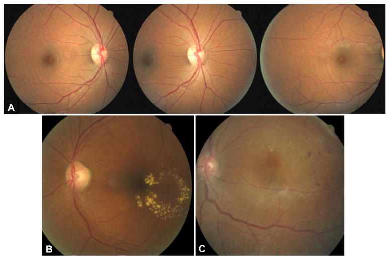
Purpose: The purpose of this study was to determine whether hard exudates (HEs) within one disc diameter of the foveola is an acceptable criterion for the referral of diabetic patients suspected of clinically significant macular edema (CSME) in a screening setting.
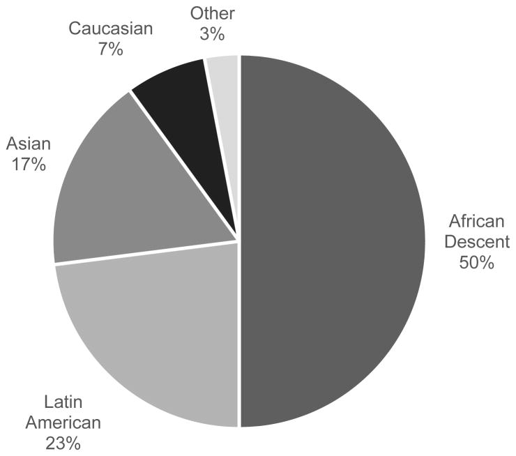
Methods: One hundred forty-three adults diagnosed as having diabetes mellitus were imaged using a nonmydriatic digital fundus camera at the Alameda County Medical Center in Oakland, CA. Nonstereo fundus images were graded independently for the presence of HE near the center of the macula by two graders according to the EyePACS grading protocol. The patients also received a dilated fundus examination on a separate visit. Clinically significant macular edema was determined during the dilated fundus examination using the criteria set forth by the Early Treatment Diabetic Retinopathy Study. Subsequently, the sensitivity and specificity of HEs within one disc diameter of the foveola in nonstereo digital images used as a surrogate for the detection of CSME diagnosed by live fundus examination were calculated.
Results: The mean (±SD) age of 103 patients included in the analysis was 56 ± 17 years. Clinically significant macular edema was diagnosed in 15.5% of eyes during the dilated examination. For the right eyes, the sensitivity of HEs within one disc diameter from the foveola as a surrogate for detecting CSME was 93.8% for each of the graders; the specificity values were 88.5 and 85.1%. For the left eyes, the sensitivity values were 93.8 and 75% for each of the two graders, respectively; the specificity was 87.4% for both graders.
Conclusions: This study supports the use of HE within a disc diameter of the center of the macula in nonstereo digital images for CSME detection in a screening setting.
Figures
Similar articles
-
Improving Accuracy of Grading and Referral of Diabetic Macular Edema Using Location and Extent of Hard Exudates in Retinal Photography.J Diabetes Sci Technol. 2015 Nov 17;10(2):262-70. doi: 10.1177/1932296815617281. J Diabetes Sci Technol. 2015. PMID: 26581880 Free PMC article.
-
High-resolution stereoscopic digital fundus photography versus contact lens biomicroscopy for the detection of clinically significant macular edema.Ophthalmology. 2002 Feb;109(2):267-74. doi: 10.1016/s0161-6420(01)00933-2. Ophthalmology. 2002. PMID: 11825807
-
A Revised Approach for the Detection of Sight-Threatening Diabetic Macular Edema.JAMA Ophthalmol. 2017 Jan 1;135(1):62-68. doi: 10.1001/jamaophthalmol.2016.4772. JAMA Ophthalmol. 2017. PMID: 27930756
-
Semi-automated quantification of hard exudates in colour fundus photographs diagnosed with diabetic retinopathy.BMC Ophthalmol. 2017 Sep 20;17(1):172. doi: 10.1186/s12886-017-0563-7. BMC Ophthalmol. 2017. PMID: 28931389 Free PMC article.
-
Application of different imaging modalities for diagnosis of Diabetic Macular Edema: A review.Comput Biol Med. 2015 Nov 1;66:295-315. doi: 10.1016/j.compbiomed.2015.09.012. Epub 2015 Sep 25. Comput Biol Med. 2015. PMID: 26453760 Review.
Cited by
-
Endothelial to mesenchymal cell transition in diabetic retinopathy: targets and therapeutics.Front Ophthalmol (Lausanne). 2023 Sep 7;3:1230581. doi: 10.3389/fopht.2023.1230581. eCollection 2023. Front Ophthalmol (Lausanne). 2023. PMID: 38983088 Free PMC article. Review.
-
Synergistic AI-resident approach achieves superior diagnostic accuracy in tertiary ophthalmic care for glaucoma and retinal disease.Front Ophthalmol (Lausanne). 2025 May 19;5:1581212. doi: 10.3389/fopht.2025.1581212. eCollection 2025. Front Ophthalmol (Lausanne). 2025. PMID: 40458618 Free PMC article.
-
Follow-up in a point-of-care diabetic retinopathy program in Pittsburgh: a non-concurrent retrospective cohort study.BMC Ophthalmol. 2024 Aug 20;24(1):356. doi: 10.1186/s12886-024-03581-9. BMC Ophthalmol. 2024. PMID: 39164678 Free PMC article.
-
Telemedicine and Diabetic Retinopathy: Review of Published Screening Programs.J Endocrinol Diabetes. 2015;2(4):10.15226/2374-6890/2/4/00131. doi: 10.15226/2374-6890/2/4/00131. Epub 2015 Nov 11. J Endocrinol Diabetes. 2015. PMID: 27430019 Free PMC article.
-
Central Macular Thickness in Diabetic Patients: A Sex-based Analysis.Optom Vis Sci. 2019 Apr;96(4):266-275. doi: 10.1097/OPX.0000000000001363. Optom Vis Sci. 2019. PMID: 30907864 Free PMC article.
References
-
- International Diabetes Federation. Clinical Guidelines Task Force. [Accessed January 3, 2014];Global Guideline for Type 2 diabetes. 2005 Available at: http://www.idf.org/webdata/docs/IDF%20GGT2D.pdf.
-
- International Society for Pediatric and Adolescent Diabetes (ISPAD), International Diabetes Federation (IDF) Global IDF/ISPAD Guideline for Diabetes in Childhood and Adolescence. International Diabetes Federation; 2011. [Accessed January 3, 2014]. Available at: http://www.idf.org/sites/default/files/Diabetes-in-Childhood-and-Adolesc....
-
- Hazin R, Barazi MK, Summerfield M. Challenges to establishing nationwide diabetic retinopathy screening programs. Curr Opin Ophthalmol. 2011;22:174–9. - PubMed
-
- Brechner RJ, Cowie CC, Howie LJ, Herman WH, Will JC, Harris MI. Ophthalmic examination among adults with diagnosed diabetes mellitus. JAMA. 1993;270:1714–8. - PubMed
-
- Han Y, Schneck ME, Bearse MA, Jr, Barez S, Jacobsen CH, Jewell NP, Adams AJ. Formulation and evaluation of a predictive model to identify the sites of future diabetic retinopathy. Invest Ophthalmol Vis Sci. 2004;45:4106–12. - PubMed
Publication types
MeSH terms
Grants and funding
LinkOut - more resources
Full Text Sources
Other Literature Sources
Medical
