

The heart as an endocrine organ
- PMID: 24562677
- PMCID: PMC3987289
- DOI: 10.1530/EC-14-0012
The heart as an endocrine organ
Abstract
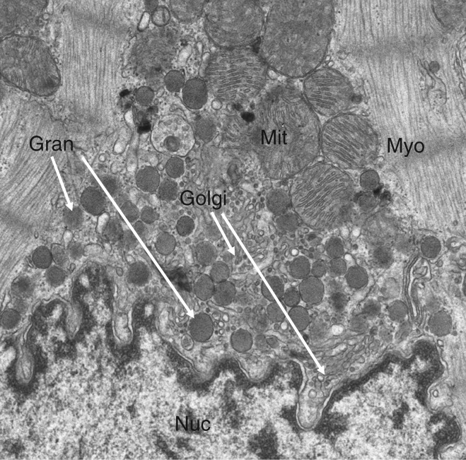
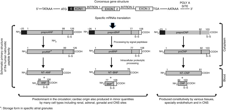
The concept of the heart as an endocrine organ arises from the observation that the atrial cardiomyocytes in the mammalian heart display a phenotype that is partly that of endocrine cells. Investigations carried out between 1971 and 1983 characterised, by virtue of its natriuretic properties, a polypeptide referred to atrial natriuretic factor (ANF). Another polypeptide isolated from brain in 1988, brain natriuretic peptide (BNP), was subsequently characterised as a second hormone produced by the mammalian heart atria. These peptides were associated with the maintenance of extracellular fluid volume and blood pressure. Later work demonstrated a plethora of other properties for ANF and BNP, now designated cardiac natriuretic peptides (cNPs). In addition to the cNPs, other polypeptide hormones are expressed in the heart that likely act upon the myocardium in a paracrine or autocrine fashion. These include the C-type natriuretic peptide, adrenomedullin, proadrenomedullin N-terminal peptide and endothelin-1. Expression and secretion of ANF and BNP are increased in various cardiovascular pathologies and their levels in blood are used in the diagnosis and prognosis of cardiovascular disease. In addition, therapeutic uses for these peptides or related substances have been found. In all, the discovery of the endocrine heart provided a shift from the classical functional paradigm of the heart that regarded this organ solely as a blood pump to one that regards this organ as self-regulating its workload humorally and that also influences the function of several other organs that control cardiovascular function.
Figures
References
-
- Kisch B. A significant electron microscopic difference between the atria and the ventricles of the mammalian heart. Experimental Medicine and Surgery. 1963;21:193–221. - PubMed
-
- de Bold AJ, Bencosme SA. Selective light microscopic demonstration of the specific granulation of the rat atrial myocardium by lead-hematoxylin-tartrazine. Stain Technology. 1975;50:203–205. - PubMed
LinkOut - more resources
Full Text Sources
Other Literature Sources
