. 2014 Apr;13(4):494-503.
doi: 10.1128/EC.00031-14. Epub 2014 Feb 21.

The Entamoeba histolytica Dnmt2 homolog (Ehmeth) confers resistance to nitrosative stress

Affiliations

The Entamoeba histolytica Dnmt2 homolog (Ehmeth) confers resistance to nitrosative stress

Rivi Hertz et al. Eukaryot Cell. 2014 Apr.

Abstract

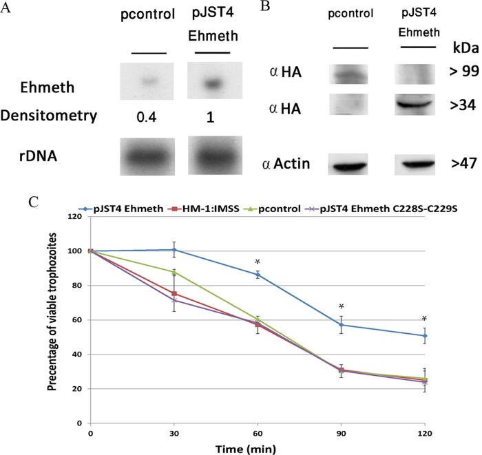
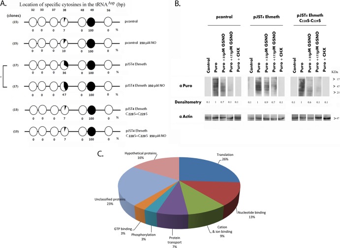
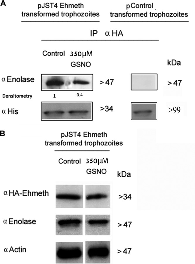
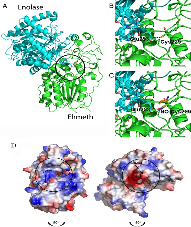
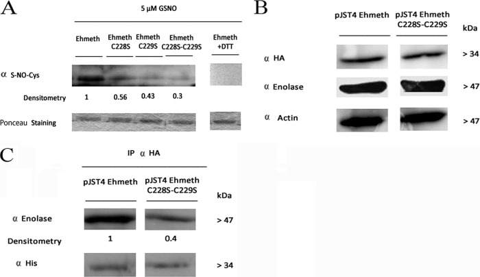
Nitric oxide (NO) has antimicrobial properties against many pathogens due to its reactivity as an S-nitrosylating agent. It inhibits many of the key enzymes that are involved in the metabolism and virulence of the parasite Entamoeba histolytica through S-nitrosylation of essential cysteine residues. Very little information is available on the mechanism of resistance to NO by pathogens in general and by this parasite in particular. Here, we report that exposure of the parasites to S-nitrosoglutathione (GSNO), an NO donor molecule, strongly reduces their viability and protein synthesis. However, the deleterious effects of NO were significantly reduced in trophozoites overexpressing Ehmeth, the cytosine-5 methyltransferase of the Dnmt2 family. Since these trophozoites also exhibited high levels of tRNA(Asp) methylation, the high levels suggested that Ehmeth-mediated tRNA(Asp) methylation is part of the resistance mechanism to NO. We previously reported that enolase, another glycolytic enzyme, binds to Ehmeth and inhibits its activity. We observed that the amount of Ehmeth-enolase complex was significantly reduced in GSNO-treated E. histolytica, which explains the aforementioned increase of tRNA methylation. Specifically, we demonstrated via site-directed mutagenesis that cysteine residues 228 and 229 of Ehmeth are susceptible to S-nitrosylation and are crucial for Ehmeth binding to enolase and for Ehmeth-mediated resistance to NO. These results indicate that Ehmeth has a central role in the response of the parasite to NO, and they contribute to the growing evidence that NO is a regulator of epigenetic mechanisms.


Figures

FIG 1
FIG 1
Overexpression of Ehmeth protects E. histolytica against nitrosative stress. (A) Northern blot analysis was performed using total RNA that was extracted from pJST4-Ehmeth and pcontrol E. histolytica trophozoites. rDNA whose expression was not changed in pJST4-Ehmeth and pcontrol trophozoites were used as controls for RNA loading. The figure displays a representative result from three independent experiments. (B) Western blot analysis was performed on nuclear protein fractions that were prepared from pJST4-Ehmeth and pcontrol E. histolytica trophozoites. The proteins were separated on 12% SDS-PAGE gels and analyzed by Western blotting with an anti-HA (α HA) antibody or an anti-actin antibody. The figure displays a representative result from three independent experiments. (C) The viabilities of wild-type E. histolytica trophozoites from strain HM-1:MSS, E. histolytica trophozoites from a strain that was transfected with a control vector (pcontrol), E. histolytica trophozoites that overexpressed Ehmeth (pJST4-Ehmeth), and E. histolytica trophozoites that overexpressed pJST4-Ehmeth C228S-C229S exposed to 350 μM GSNO for 30, 60, 90, and 120 min were measured. The number of trophozoites at the beginning of each experiment was set at 100%. Bars represent the standard deviations of the means. The means of the different groups for three independent experiments were compared using Student's t test, and statistical significance was set at 5%. The viabilities of the wild-type E. histolytica trophozoites of strain HM-1:MSS, the pcontrol E. histolytica trophozoites, and the pJST4-Ehmeth C228S-C229S E. histolytica trophozoites were not significantly different from each other at any time point, In contrast, the viability of the pJST4-Ehmeth E. histolytica trophozoites was significantly different (P < 0.05) from that of the wild-type E. histolytica trophozoites of strain HM-1:MSS, the pcontrol E. histolytica trophozoites, and the pJST4-Ehmeth C228S-C229S E. histolytica trophozoites after a 60- or 120-min exposure to GSNO.
FIG 2
FIG 2
Level of tRNAAsp methylation in NO-treated trophozoites. (A) Bisulfite sequencing analysis of tRNAAsp in pcontrol E. histolytica trophozoites, pcontrol E. histolytica trophozoites that were treated with 350 μM GSNO for 1 h, pJST4-Ehmeth trophozoites, pJST4 Ehmeth trophozoites treated with GSNO (350 μM for 1 h), pJST4-Ehmeth C228S-C229S trophozoites, and pJST4-Ehmeth C228S-C229S trophozoites treated with GSNO (350 μM for 1 h). The numbers of clones (sequence reads) are displayed in parentheses on the left side of each row. Black areas in the circles indicate methylated cytosine residues, and white areas indicate unmethylated cytosine residues. The percentage of methylated cytosines is indicated below each circle. The location of specific cytosines in the tRNAAsp is indicated under each row. The levels of C38 tRNAAsp methylation in the untreated pcontrol and GSNO-treated pcontrol trophozoites were not significantly different from each other according to the analysis with Student's t test, for which statistical significance was set at 5%. In contrast, the levels of C38 tRNAAsp methylation in the untreated and GSNO-treated pJST4 Ehmeth trophozoites were significantly different (P < 0.05). (B) Protein synthesis, measured using puromycin-labeled proteins. pcontrol E. histolytica trophozoites, pJST4-Ehmeth E. histolytica trophozoites, and pJST4-Ehmeth C228S-C229S E. histolytica trophozoites were mock treated (control), labeled with 10 μg/ml puromycin (Puro), or treated with 35 μM or 175 μM GSNO for 15 min and then labeled with puromycin for 20 min (Puro + GSNO). Some trophozoites were treated with cycloheximide (CHX; 100 μg/ml) before puromycin labeling (Puro + CHX). The extracts were separated by denaturing electrophoresis and analyzed by Western blotting with a 12D10 clone puromycin antibody. An actin immunoblot is shown as the loading control. The results are representative of two independent experiments. α-Puro, anti-Puro antibody. (C) Functional categories of the upregulated proteins in pJST4-Ehmeth E. histolytica trophozoites and pcontrol E. histolytica trophozoites exposed to 350 μM GSNO for 1 h. The upregulated proteins were classified according to their biological role based on the David Bioinformatics Resources (http://david.abcc.ncifcrf.gov/).
FIG 3
FIG 3
Nitric oxide regulates the amount of Ehmeth-enolase inhibitory complex formed. (A) Ehmeth samples from nuclear lysates of pJST4-Ehmeth and pJST4 Ehmeth E. histolytica trophozoites that were treated with 350 μM GSNO for 1 h was immunoprecipitated (IP) with a monoclonal anti-HA (α HA) antibody. The presence of enolase among the immunoprecipitated proteins was detected by Western blotting with an enolase antibody (left). The presence of CHH-tagged Ehmeth among the immunoprecipitated proteins was detected by Western blotting by using a histidine antibody. The immunoprecipitation experiments were also performed with nuclear lysates of pcontrol E. histolytica trophozoites as a negative control for the expression of CHH-tagged Ehmeth and for the immunoprecipitation of enolase (right). (B) Western blot analysis of nuclear proteins prepared from GSNO-treated pJST4-Ehmeth and pJST4-Ehmeth E. histolytica trophozoites. The proteins were separated on 12% SDS-PAGE gels and analyzed by Western blotting with an HA antibody, an enolase antibody, or an actin antibody. The figure displays a representative result from at least three independent experiments.
FIG 4
FIG 4
Molecular modeling of the putative enolase-Ehmeth complex. The atomic coordinates of E. histolytica enolase and Ehmeth proteins (PDB codes 3QTP and 3QV2, respectively) were docked using the Hex 6.2 platform. (A) The overall best docked structure. (B and C) Close-up images of the interaction between the Glu253 residue of enolase and the Cys229 residue of Ehmeth in the native (B) and NO-modified (C) forms. The complex shown in panel A was disassembled by rotating the enolase 90° in a clockwise direction and the Ehmeth 90° in a counterclockwise direction (the black arrows indicate the direction of complex formation). The interaction interfaces of both proteins are indicated by black circles. (D) Vacuum electrostatic potentials were generated using PyMOL in order to illustrate the charge variance of the E. histolytica enolase and Ehmeth proteins, with red and blue indicating negative and positive surfaces, respectively.
FIG 5
FIG 5
Role of Ehmeth Cys228 and Cys229 in the formation of the Ehmeth-enolase complex. (A) Western blot analysis of recombinant proteins (Ehmeth, Ehmeth C228S, Ehmeth C229S, and Ehmeth C228S-C229S) that were treated with 5 μM GSNO for 1 h at 37°C. The proteins were resolved on 12% polyacrylamide gels under native conditions, transferred to a nitrocellulose membrane, and then probed with an S-NO-Cys antibody (α S-NO-Cys). Ponceau staining of the membrane prior to its interaction with the S-NO-Cys antibody was used as a loading control. The Ehmeth plus DTT control shows the results with the Ehmeth recombinant protein treated with 5 μM GSNO for 1 h at 37°C followed by incubation with 20 mM DTT for 5 min at 37°C. The figure displays a representative result from at least three independent experiments performed singly. (B) Western blot analysis of nuclear protein fractions prepared from pJST4-Ehmeth and pJST4-Ehmeth C228S-C229S E. histolytica trophozoites performed using an HA antibody, an enolase antibody, or an actin antibody. The figure displays a representative result from at least three independent experiments performed singly. (C) Immunoprecipitation analysis of Ehmeth from pJST4-Ehmeth and pJST4-Ehmeth C228S-C229S E. histolytica trophozoites, performed with an HA antibody. The presence of enolase among the immunoprecipitated proteins was detected by using an enolase antibody. The amounts of Ehmeth and Ehmeth C228S-C229S in the pJST4-Ehmeth and pJST4-Ehmeth C228S-C229S E. histolytica trophozoites were determined by using a histidine antibody.

Similar articles

Cited by

References

    1. . 1997. WHO/PAHO/UNESCO report. A consultation with experts on amoebiasis. Mexico City, Mexico 28–29 January, 1997. Epidemiol. Bull. 18:13–14 - PubMed
    1. Baumel-Alterzon S, Weber C, Guillen N, Ankri S. 2013. Identification of dihydropyrimidine dehydrogenase as a virulence factor essential for the survival of Entamoeba histolytica in glucose-poor environments. Cell. Microbiol. 15:130–144. 10.1111/cmi.12036 - DOI - PubMed
    1. Tovy A, Hertz R, Siman-Tov R, Syan S, Faust D, Guillen N, Ankri S. 2011. Glucose starvation boosts Entamoeba histolytica virulence. PLoS Negl. Trop. Dis. 5(8):e1247. 10.1371/journal.pntd.0001247 - DOI - PMC - PubMed
    1. Mortimer L, Chadee K. 2010. The immunopathogenesis of Entamoeba histolytica. Exp. Parasitol. 126:366–380. 10.1016/j.exppara.2010.03.005 - DOI - PubMed
    1. Wolfe AJ. 2005. The acetate switch. Microbiol. Mol. Biol. Rev. 69:12–50. 10.1128/MMBR.69.1.12-50.2005 - DOI - PMC - PubMed

Publication types

MeSH terms

LinkOut - more resources